Quantcast
Channel: AnatomyZone
Viewing all 279 articles
Browse latest View live

External Abdominal Oblique Muscle
$
0
0

External Abdominal Oblique Muscle

External Abdominal Oblique Muscle

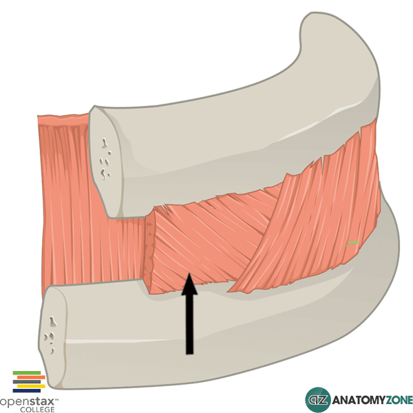
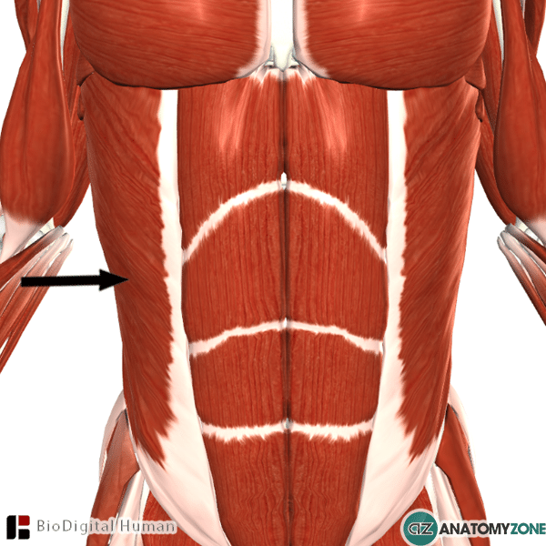
The muscle indicated is the external oblique muscle of the abdomen.

The external oblique muscle is the most superficial lateral muscle of the anterior abdominal wall. The three muscles which make up the lateral part of the anterior wall of the abdomen from superficial to deep are the:

  • External oblique
  • Internal oblique
  • Transversus abdominis

Origin: ribs 5-12

Insertion: Iliac crest, pubic tubercle, linea alba

Innervation: thoracoabdominal nerve and subcostal nerve

Action: Flexion and rotation of the vertebral column – brings about contralateral rotation of the torso. Unilateral contraction causes lateral flexion of the vertebral column. Important in increasing intra-abdominal pressure.

The aponeurosis of the external oblique muscle is important, as it forms the inguinal ligament, which runs from the pubic tubercle to the anterior superior iliac spine. The ligament forms the base of the inguinal canal.

The fibres of the external abdominal oblique muscle are oriented inferiorly and anteriorly – the fibres of the internal oblique are oriented perpendicular, running superomedially.

Learn more about the anatomy of the external oblique muscle in this tutorial on the anterior abdominal wall.

Longitudinal Muscle Layer of Stomach

$
0
0

Longitudinal Muscle Layer of Stomach

Longitudinal Muscle Layer of Stomach

The structure indicated is the outer longitudinal muscle layer of the stomach.

The stomach walls consists of four parts:

  • Mucosa
  • Submucosa
  • Muscularis externa
  • Serosa

The muscularis externa is the muscular layer of the gastrointestinal wall. In the stomach it consists of three layers:

  • Inner oblique layer
  • Middle circular layer
  • Outer longitudinal layer

Between the middle circular layer and outer longitudinal muscular layer lies Auerbach’s plexus (the myenteric plexus).

Learn more about the digestive system in this tutorial

Superior Pubic Ramus

$
0
0

Superior Pubic Ramus

Superior Pubic Ramus

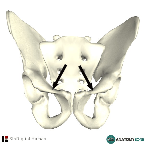
The structure indicated is the superior pubic ramus.

The pelvic bone consists of three components:

  • Ilium
  • Pubis
  • Ischium

The pubis is the anterior and inferior part of the pubic bone and consists of a body connected to two branches (rami) – a superior ramus and an inferior ramus. Several muscles of the abdomen and thigh attach to the superior pubic ramus.

The superior pubic ramus projects posteriorly and laterally from the body and joins the ilium and ischium. The superior pubic ramus consists of two parts: a medial part (the body of the pubis) and a lateral part. The medial part consists of two surfaces (anterior, posterior) and three borders (upper, medial, lateral). The lateral part consists of three surfaces (superior, inferior, posterior).

The pectineal line (pecten pubis) is a ridge on top of the superior ramus. Combined with the arcuate line, this forms the iliopectineal line. Anteriorly, this line is continuous with the pubic crest.

Learn more about the anatomy of the bones of the pelvis in this tutorial

Styloglossus Muscle

$
0
0

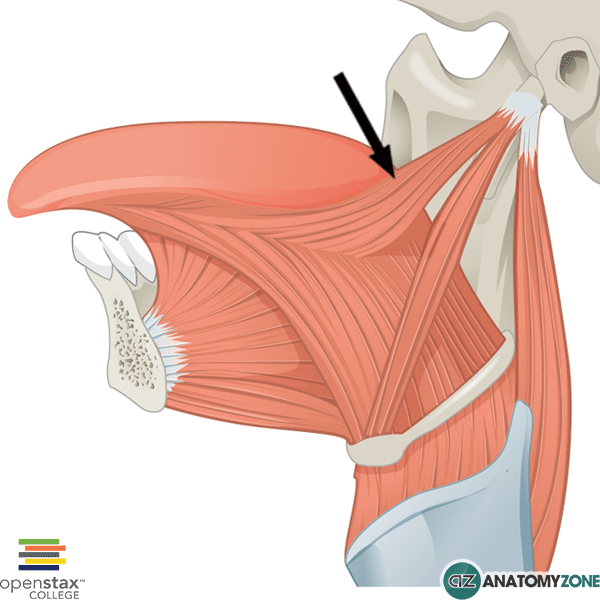
Styloglossus Muscle

Styloglossus Muscle

The muscle indicated is the styloglossus muscle       .

The styloglossus muscles is one of three styloid muscles. The three styloid muscles are:

  • Styloglossus
  • Stylohyoid
  • Stylopharyngeus

Origin: Styloid process of temporal bone

Insertion: lateral aspect of tongue

Innervation: Hypoglossal nerve (cranial nerve 12)

Action: Elevation and retraction of the tongue

The styloid muscle is one of several extrinsic muscles of the tongue.

Learn all about the extrinsic muscle of the tongue in this anatomy tutorial.

Internal Intercostal Muscle

$
0
0

Internal Intercostal Muscle

Internal Intercostal Muscle

The structure indicated is the internal intercostal muscle.

The intercostal muscles are a set of three flat muscles which are located in each intercostal space. The three intercostal muscles, from superficial to deep are:

  • External intercostal
  • Internal intercostal
  • Innermost intercostal

Origin: Lateral edge of costal groove of rib directly above

Insertion: Superiorly on rib directly below

Innervation: Intercostal nerves (T1 – T11)

Action: Forced expiration, movement of ribs inferiorly

The muscle fibres of the internal intercostal muscles pass obliquely in the opposite direction to the external intercostal muscles, whose fibres pass anteroinferiorly (when the thoracic wall is viewed in the lateral position).

Learn more about the muscles of the thoracic wall in this anatomy tutorial.

Abducent Nerve

$
0
0

Abducent Nerve

Abducent Nerve
The structure indicated is the abducent nerve (cranial nerve VI).

The abducent nerve (cranial nerve VI), provides general somatic efferent fibres (motor function) to the lateral rectus muscle of the eye. There are three cranial nerves which are responsible for innervating the extrinsic muscles of the eye:

  • Oculomotor nerve (cranial nerve III)
  • Trochlear nerve (cranial nerve VI)
  • Abducent nerve

The oculomotor nerve supplies the superior, inferior and medial rectus muscles, as well as the inferior oblique muscle and the levator palpebrae superioris.

The trochlear nerve supplies the superior oblique muscle.

It originates between the pons and the medulla of the brain stem. It has a long intracranial course which makes it vulnerable to injury: fractures of the petrous part of the temporal bone, aneurysms of the internal carotid artery and mass lesions all put this nerve at risk. Lesions of the abducent nerve cause diplopia on lateral gaze, with inability to abduct the affected eye – the eye is pulled medially due to the unopposed action of the medial rectus muscle.

Learn more about the cranial nerves in this anatomy tutorial

Left Anterior Descending Artery

$
0
0

Left Anterior Descending Artery

Left Anterior Descending Artery

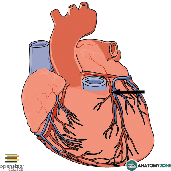
The structure indicated is the left anterior descending artery (also known as the anterior interventricular artery).

The left anterior descending artery is one of the coronary arteries which supplies the anterior and lateral parts of the myocardium and the interventricular septum. It is responsible for approximately 45-55% of the left ventricles blood supply.

The left coronary artery arises from the left aortic sinus of the ascending aorta. The left coronary artery has two terminal branches:

  • Left anterior descending (LAD)/anterior interventricular
  • Circumflex branch

The left anterior descending artery itself has two different types of branches

  • Septal branches
  • Diagonal branches

Septal branches supply the anterior two thirds of the interventricular septum.

Diagonal branches supply the lateral wall of the left ventricle as well as the anterior papillary muscle.

This artery is often referred to morbidly as the “widow maker” – occlusion of the LAD can have catastrophic consequences due to the large proportion of blood it supplies to the left ventricle.

Learn more about the arterial supply to the heart in this anatomy tutorial.

Navicular Bone

$
0
0

Navicular Bone

Navicular Bone

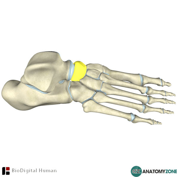
The structure highlighted is the navicular bone of the foot.

The foot contains three groups of bones:

  1. Tarsals
  2. Metatarsals
  3. Phalanges

The navicular is one of seven tarsal bones. There are three groups of tarsal bones:

  1. Proximal group
  2. Intermediate group
  3. Distal group

The navicular bone is the only bone in the intermediate group and is located medially in the foot. Posteriorly, the navicular articulates with the talus. Anteriorly, the navicular articulates with the cuneiform bones and laterally it articulates with the cuboid bone.

There is only one muscle which attaches to the navicular bone: the tibialis posterior. It attaches onto the tuberosity of the navicular bone.

Learn more about the bones of the foot in this anatomy tutorial.


Capitate Bone

$
0
0

Capitate Bone

Capitate Bone

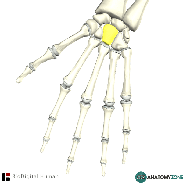
The structure highlighted is the capitate bone of the hand.

There are three groups of bones in the hand:

  • Carpal bones (8 in total)
  • Metacarpal bones
  • Phalanges

The carpal bones are separated into two rows:

  • Proximal row
  • Distal row

The capitate bone is located in the distal row of carpal bones. It is the largest of the carpal bones and is located centrally in the wrist. The capitate bone has a “head” which articulates with the scaphoid and lunate bones of the proximal row.

Laterally, on the radial side is the trapezoid bone, on medially, on the ulnar side is the hamate bone.

The capitate bone has 6 surfaces

  • Dorsal
  • Palmar
  • Superior
  • Inferior
  • Medial
  • Lateral

Learn more about the bones of the hand in this anatomy tutorial.

Quadratus Plantae Muscle

$
0
0

Quadratus Plantae Muscle

Quadratus Plantae Muscle

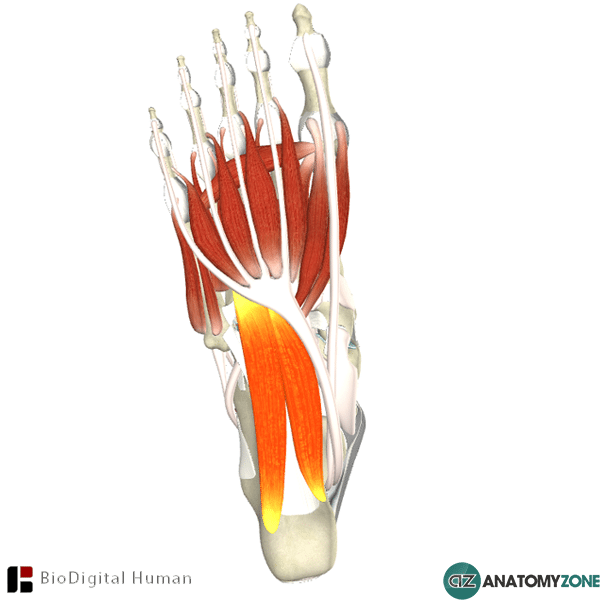
The muscle highlighted is the quadratus plantae muscle of the foot.

The intrinsic muscles on the plantar aspect of the foot are divided into four layers. The quadratus plantae muscle is found in the second layer of muscles. There are two muscles in the second layer:

  • Quadratus plantae
  • Lumbricals

The muscles in the second layer are associated with the tendons of the flexor digitorum longus muscle. This tendon forms the site of insertion for the quadratus plantae muscle, and forms the site of origin for the four lumbricals. The quadratus plantae muscle has two heads – a medial head and a lateral head.

Origin: Medial surface of calcaneus and lateral process of calcaneal tuberosity

Insertion: Lateral aspect of flexor digitorum longus tendon.

Innervation: Lateral plantar nerve

Action: Flexion of toes 2-5 (assists the action of flexor digitorum longus)

Learn more about the anatomy of the muscles of the foot in this tutorial.

Right Subclavian Artery

$
0
0

Right Subclavian Artery

Right Subclavian Artery

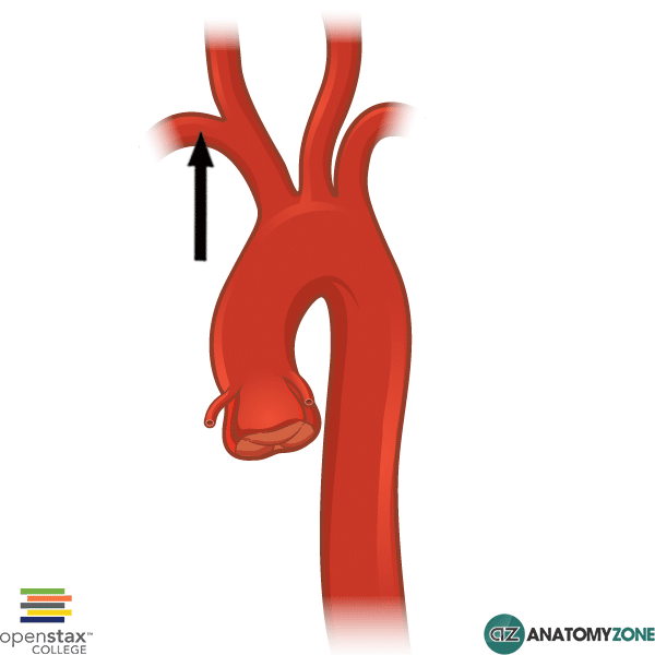
The structure indicated is the right subclavian artery.

The subclavian arteries are located under (“sub”) the clavicle (“clavian”) and receive blood from the aortic arch. The subclavian arteries supply the arms, with some branches that extend to supply the head.

The branches of the aortic arch are:

  • Brachiocephalic artery
  • Left common carotid artery
  • Left subclavian artery

The left subclavian artery comes directly off the arch of the aorta, whereas on the right side of the body, the brachiocephalic artery splits, giving rise to the right subclavian artery, and the right common carotid artery.

The subclavian artery becomes the axillary artery at the lateral border of the first rib. It can be thought of in three parts, and gives rise to several branches:

  • First part: vertebral artery, internal thoracic artery, thyrocervical trunk
  • Second part: costocervical trunk
  • Third part: dorsal scapular artery

Learn more about the heart and the great vessels in this anatomy tutorial.

Capsule of Lymph Node

$
0
0

Capsule of Lymph Node

Capsule of Lymph Node

This picture represents a cross section of a lymph node. The arrow indicates the capsule of the lymph node.

Lymph nodes are organs of the lymphatic system located in several areas throughout the body, providing the major site of immune cells, including B and T cells. The lymph node is surrounded by a fibrous capsule, which projects internally to form trabeculae. Lymph nodes consist of an outer cortex and an inner medulla.

From superficial to deep, the lymph node consist of the following components:

  • Capsule
  • Subcapsular sinus
  • Cortex
  • Medulla

The capsule of the lymph node consists of dense irregular connective tissue, and from this capsule several trabeculae extend internally. Between the outer cortex and the fibrous capsule is the subcapsular sinus. Lymph flows through afferent lymphatic vessels into the subcapsular sinuses, which then drains into trabecular sinuses and then the medullary sinuses.

Pancreatic Duct

$
0
0

Pancreatic Duct

Pancreatic Duct

The structure indicated is the pancreatic duct (duct of Wirsung) of the pancreas.

The pancreas is an organ which lies posteriorly to the stomach and extends from the duodenum to the spleen.

The pancreatic duct extends from the tail of the pancreas through the body of the pancreas into the head of the pancreas where it joins the common bile duct to form the hepatopancreatic ampulla, also known as the ampulla of Vater, which empties into the descending duodenum at the major duodenal papilla.

In addition to the main pancreatic duct, there is also an accessory pancreatic duct (duct of Santorini), which empties into the minor duodenal papilla.

Obstruction of the pancreatic duct, commonly due to gallstones can lead to acute pancreatitis.

Learn more about the basic anatomy of the organs of the digestive system in this tutorial.

Coccyx

$
0
0

Coccyx

Coccyx

The structure indicated is the coccyx.

The coccyx forms the terminal aspect of the spinal column, and is comprised of three to five fused vertebrae. In humans and other great apes, the coccyx is the remnant of a vestigial tail. Superiorly, the coccyx articulates with the sacrum. On this superior surface is a facet for articulation with the sacrum, and also two processes which extend superiorly, known as cornua or horns.

On the lateral surface of the coccygeal vertebrae are transverse processes – this process is most prominent on the first coccygeal vertebrae. The coccyx contains no spinous processes, pedicles or laminae.

Coccygodynia as a painful condition of the coccyx, often following sudden impact on the coccyx following trauma from a fall, or from the pressure on the coccyx during childbirth in women.

Gray Ramus Communicans

$
0
0

Gray Ramus Communicans

Gray Ramus Communicans

The structure indicated is the gray ramus communicans (known collectively as the gray rami communicantes).

The spinal nerves at each level receive this branch, the gray ramus communicans, conveying postganglionic nerve fibres from the sympathetic nervous system. In the same picture, parallel to the gray ramus communicans, the branch which conveys the preganglionic sympathetic fibres into the paravertebral sympathetic ganglia is known as the white ramus communicans.

The preganglionic nerve fibre which passes through the white ramus communicans has three potential outcomes:

  1. Synapse in the paravertebral ganglion.
  2. Travel up or down a level in the sympathetic chain to synapse with a paravertebral ganglion at a higher or lower level.
  3. Travel straight through the paravertebral ganglion without forming a synapse, to form a synapse at a distant ganglion anterior to the vertebral column, in a plexus.

Learn more about the structure of the sympathetic nervous system in this anatomy tutorial.


Tibialis Anterior

$
0
0

Tibialis Anterior

Tibialis Anterior

The muscle highlighted is the tibialis anterior muscle.

The tibialis anterior is one of the four muscles of the anterior compartment of the leg. The muscles of the leg consist of three compartments:

  • Anterior
  • Posterior
  • Lateral

In the anterior compartment are the four following muscles:

  • Tibialis anterior
  • Extensor hallucis longus
  • Extensor digitorum longus
  • Fibularis tertius

The muscles of the anterior compartment of the leg serve to dorsiflex the ankle joint, extend the toes and invert the foot, and they are supplied by the deep fibular (peroneal) nerve.

Origin: Upper two-thirds and lateral condyle of the tibia

Insertion: Medial cuneiform and first metatarsal of the foot

Innervation: Deep fibular nerve

Action: Dorsiflexion and inversion of the foot

Learn more about the anatomy of the leg muscles in this tutorial!

Carpal Tunnel

$
0
0

Carpal Tunnel

Carpal Tunnel

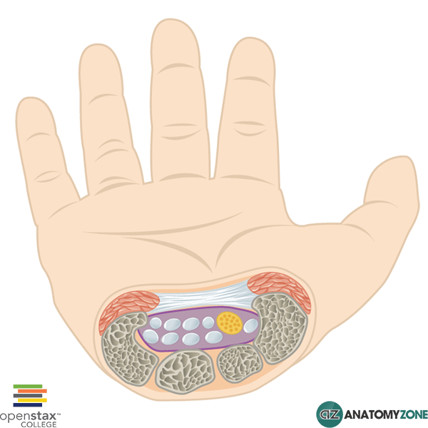
Represented in this diagram is the carpal tunnel.

The carpal tunnel is formed by an arch of carpal bones and the flexor retinaculum, found on the anterior aspect of the wrist. The flexor retinaculum attaches on the radial side to the scaphoid tubercle and the trapezium bone; on the ulnar side it attaches to the hook of the hamate and the pisiform. Its attachment over the arch of carpal bones converts the space into a tunnel which allows for the passage of several tendons and the median nerve.

The carpal tunnel contains nine long flexor tendons and the median nerve:

  • Flexor digitorum profundus (four tendons)
  • Flexor digitorum superficialis (four tendons)
  • Flexor pollicis longus (one tendon)

The tendon of flexor carpi radialis passes through the flexor retinaculum in its own compartment – it does not technically pass through the carpal tunnel.

Carpal tunnel syndrome occurs due to pressure on the median nerve within the carpal tunnel. This typically causes sensations of pain/pins and needles in the distribution of the median nerve, as well as muscle weakness and atrophy of the thenar muscles.

Common causes of carpal tunnel syndrome include rheumatoid arthritis, pregnancy, hypothyroidism, acromegaly and trauma.

Styloid Process of Radius

$
0
0

Styloid Process of Radius

Styloid Process of Radius

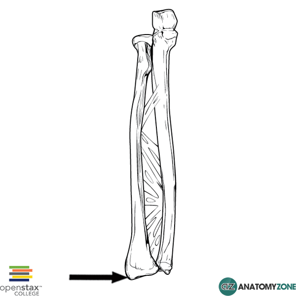
The structure indicated in this diagram is the styloid process of the radius.

The distal end of the radius consists of:

  • Radial styloid process
  • Two facets for articulation with the scaphoid and lunate bone
  • Ulnar notch

The radial styloid process projects obliquely downward from the distal end of the radius. It serves as the point of attachment for the brachioradialis muscle and the radial collateral ligament.

Laterally, the tendons of extensor pollicis brevis and abductor pollicis longus run in a flat groove.

Chauffeur’s fracture is the name given to fracture of the radial styloid and typically occurs when the scaphoid bone is compressed against the styloid process – often due to falling onto an outstretched hand.

Learn more about the anatomy of the radius and ulnar in this tutorial, and about the anatomy of the wrist in this tutorial.

Lateral Semicircular Canal

$
0
0

Lateral Semicircular Canal

Lateral Semicircular Canal

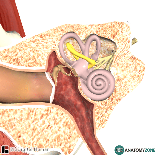
The structure highlighted is the lateral semicircular canal.

The ear consists of three anatomical components:

  • External ear
  • Middle ear
  • Internal ear

The internal ear consists of the bony labyrinth and the membranous labyrinth. The semicular canals are a component of the bony labyrinth, which consists also of the vestibule and the cochlea. These bony components contain a clear fluid known as perilymph.

There are three semicircular canals:

  • Anterior (superior) semicircular canal
  • Posterior semicircular canal
  • Lateral (horizontal) semicircular canal

Each semicircular canal attaches to the vestibule and has one end that is enlarged to form the ampulla. The semicircular canals are oriented at right angles to each other – these different orientations allow for individual canals to be stimulated according to rotation and movement of the head in different planes.

The lateral semicircular canal is the shortest of the canals and is responsible for conveying rotational motion of the head around the vertical axis. This is the movement that is produced if, when sitting upright, you were to move your head from left to right, and vice versa.

Sacrotuberous Ligament

$
0
0

Sacrotuberous Ligament

Sacrotuberous Ligament

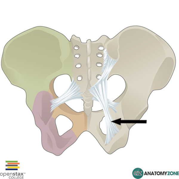
The structure indicated is the sacrotuberous ligament.

The sacrotuberous ligament runs from the sacrum (sacro-) to the ischial tuberosity (-tuberous). The sacrotuberous ligament contains the coccygeal branch of the inferior gluteal artery. In combination with the sacrospinous ligament, the two ligaments serve to form two important foramina which allow structures to exit the pelvis:

  • Greater sciatic foramen
  • Lesser sciatic foramen

The sacrospinous ligament runs from the sacrum and coccyx to the ischial spine. The greater sciatic foramen is formed by the sacrospinous ligament and the greater sciatic notch; the lesser sciatic foramen is formed by the sacrotuberous ligament and the lesser sciatic notch.

Sometimes the pudendal nerve can become trapped between the sacrotuberous ligament and the sacrospinous ligament, causing perineal pain (pudendal nerve entrapment/pudendal neuralgia).

Learn all about the anatomy of the pelvis in this tutorial.

Viewing all 279 articles
Browse latest View live