Part 1 – Testis and Epididymis
Part 2 – Vas Deferens, Accessory Glands and Urethra
Part 3 – The Penis
About this video
The renal pelvis is the proximal dilated aspect of the ureter in the kidney. It functions to funnel urine into the ureters.
Urine empties from the renal papilla into the minor calyces, which in turn drain into the major calyces, and then into the renal pelvis.
To learn more about the internal anatomy of the renal pelvis take a look at this video.
The biceps brachii is a muscle in the anterior compartment of the arm. It has two heads, the long head and the short head. It is one of three muscles in the anterior compartment of the arm, the other two muscles being the brachialis and the coracobrachialis, which lie deep to it.
Despite being a relatively small muscle, the biceps brachii is one of the most highly trained muscles in the gym, along with the pectoral muscles and the deltoid muscles.
If you are a fitness enthusiast, what exercises do you use to train this muscle?
Learn about the muscles of the anterior compartment in this video and check out our 3D atlas where you can visualise and interact with these muscles for yourself.
Muscle | Action | Origin | Insertion | Innervation | Blood supply |
---|---|---|---|---|---|
Biceps Brachii | - Flexion of elbow - Supination of forearm | - Short head: Coracoid process - Long head: Supraglenoid Fossa | - Radial tuberosity | - Musculocutaneous nerve | - Brachial artery |
Brachialis | - Flexion of elbow | - Distal half of anterior humerus | - Ulna tuberosity | - Musculocutaneous nerve | - Brachial artery |
Coracobrachialis | - Assists in flexion of shoulder | - Coracoid Process | - Medial midshaft humerus | - Musculocutaneous nerve | - Brachial artery |
The inferior colliculi together with the superior colliculi, are collectively referred to as the corpora quadrigemina and form the roof of the midbrain.
The word colliculus actually means “lower hill” in Latin, describing the appearance of these structures.
The superior colliculi are part of the visual system, whereas the inferior colliculi are involved in the auditory system.
Auditory fibres project to the inferior colliculi, which then in turn project to the medial geniculate nucleus of the thalamus. From here, the fibres project to the auditory cortex within the temporal lobe.
Learn about the basic anatomy of the brain in this video.
The semitendinosus muscle is one of three muscles in the posterior compartment of the thigh. The other muscles are the semimembranosus, and the biceps femoris.
The muscles in the posterior compartment are often referred to as the “hamstrings” muscles. Collectively, these muscles are responsible for extending the hip joint, and flexing the knee joint.
The biceps femoris lies laterally, and the semitendinosus and semimembranosus lie medially.
The semitendinosus gets its name from its structure which is comprised of a muscle belly which tapers into a long tendon that extends to insert onto the medial tibia. The semitendinosus sits on top of the semimembranosus. An easy way to remember this relationship is semitendinosus top.
You can learn more about the muscles of the posterior compartment in this video.
Check out our 3D atlas where you can visualise and interact with these muscles for yourself.
The right lung consists of three lobes:
These lobes are separated by two fissures, known as the oblique fissure and the horizontal fissure.
The oblique fissure separates the middle and inferior lobes, whereas the horizontal fissure separates the superior and middle lobes.
Pictured here is the middle lobe of the right lung.
The left lung, on the other hand only has two lobes separated by one fissure. The superior lobe is separated from the inferior lobe by the oblique fissure.
Learn more about the respiratory system in this tutorial.
The ossicles of the ear are tiny little bones that are located in the middle ear. They are the smallest bone in the human body (the word itself actually means “little bone”) and their purpose is to transmit sound vibrations from the eardrum (tympanic membrane), to the complex inner ear structures, ultimately leading to the perception of sound.
There are a series of three ossicles in the ear:
The malleus is attached to the tympanic membrane and articulates with the incus, which in turn articulates with the stapes. The stapes, attaches to the membrane of the oval window connecting the middle ear to the inner ear.
Through this series of bones, the vibrations are transmitted from the tympanic membrane to the inner ear.
The ankle joint has medial and lateral ligaments which help to keep it stable.
As the “fibular” part of the name suggests, the anterior talofibular ligament is one of the lateral ligaments. There are three ligaments in this lateral ligament complex:
This ligament is the most commonly injured ligament in a sprained ankle. Since the ligament stabilises the lateral side of the ankle, you can image from seeing its position, if the ankle is forced into inversion, this ligament will come under a lot of strain leading to a tear.
Check out our 3D atlas where you can visualise and interact with the ankle joint yourself.
The arrector pili muscles are small smooth muscles which attach to hair follicles. When these muscles contract, they make your hair stand on end – a phenomenon which may better known to you as “goosebumps”.
These muscles are not under conscious control, but are innervated by the sympathetic component of the autonomic nervous system.
Find out more about the autonomic nervous system in this tutorial.
The vastus medialis is one of the muscles that makes up the quadriceps femoris, and is contained in the anterior compartment of the thigh. It is responsible for the tear-drop shape that is visible on the medial aspect of the thigh in trained athletes and footballers – just watch the world cup and see for yourself!
The four muscles that make up the quadriceps femoris are the:
- Rectus femoris
- Vastus lateralis
- Vastus medialis
- Vastus intermedius
Learn more about the muscles of the anterior compartment in this tutorial.
Click here to visualise and interact with these muscles.
The structure indicated is the lateral rectus muscle.
This muscle is one of six muscles that are involved in the movements of the eye. The muscles which move the eye are referred to as the extraocular muscles, these include:
Origin: Lateral part of annulus of Zinn
Insertion: Lateral (temporal) aspect of eyeball
Action: Abducts the eyeball – moves it away from the midline
Innervation: Abducens nerve (Cranial nerve VI)
If the abducens nerve is damaged (abducens nerves palsy/sixth nerve palsy), there is failure to abduct the affected eyeball, causing double vision in the horizontal plane.
The arrow is pointing to the greater trochanter of the femur.
The greater trochanter is a large quadrilateral eminence which forms the site of origin and insertion for several muscles:
Origins: vastus lateralis
Insertions: gluteus medius, gluteus minimus, obturator internus, piriformis
The greater trochanter has two surfaces:
At the base of the greater trochanter is a deep depression known as the trochanteric fossa. Posteriorly is the intertrochanteric crest. The trochanteric fossa serves as the point of insertion for four muscles:
The greater trochanter has four borders:
Learn all about the muscles of the thigh in this tutorial.
Learn all about the features of the femur in this tutorial.
The structure indicated is the pulmonary valve.
There are four sets of valves in the heart. Between the atria and the ventricles are the atrioventricular valves; on the right is the tricuspid valve, on the left is the mitral (bicuspid) valve.
Between the ventricles and the aorta/pulmonary trunk are the semilunar valves. The pulmonary valve is located between the right ventricle and the pulmonary trunk, and the aortic valve is located between the left ventricle and the aorta.
The pulmonary valve (pulmonic valve) has three cusps. The pulmonary valve opens in ventricular systole, when the pressure in the right ventricle becomes greater than the pressure in the pulmonary trunk, thereby forcing the valve open. At the end of ventricular systole, the pressure in the right ventricle falls, and the pressure in the pulmonary trunk is greater, thereby forcing the closure of the pulmonary valve.
The three cusps of the pulmonary valve are:
The free superior edge of these cusps thickens in the middle to form a nodule, laterally it is thinner and is called the lunule.
Learn more about the anatomy of the heart in these tutorials:
The structure indicated is the anterior communicating artery.
The anterior communicating artery connects the left cerebral artery to the right cerebral artery. It is one of the blood vessels which contributes to the arterial Circle of Willis – an anastomosis of arterial vessels located on the base of the brain, consisting of the following arteries:
The anterior cerebral artery also gives rise to small collateral vessels which supply the frontal lobes and basal forebrain. This vessel is a common site for the formation of aneurysms. Saccular aneurysms, known as berry aneurysms are round outpouchings of the vessel wall and the anterior communicating artery is the most common site for these types of aneurysm to form. Rupture of these aneurysms leads to haemorrhage into the subarachnoid space.
Learn more about the Circle of Willis in this tutorial!
The structure indicated is the azygos vein.
The azygos system of veins includes:
The azygos vein runs in the posterior mediastinum, on the right side of thoracic vertebral column. It drains into the superior vena cava, and transports deoxygenated blood from the posterior walls of the thorax and the abdomen.
The azygos system forms important anastamotic connections, which serve to return blood to the heart via the superior vena cava, if the inferior vena cava becomes obstructed.
The structure indicated is the right main bronchus.
The trachea divides into two main bronchi – the right main bronchus and the left main bronchus (also referred to as primary bronchi). The trachea divides at the level of the sternal angle and the point at which it bifurcates is known as the carina – a cartilaginous ridge which runs between the left and right main bronchi.
The main bronchi then divide into smaller and smaller airways in the following sequence:
The right main bronchus is wider than the left main bronchus and is oriented more vertically than the left main bronchus. Due to this difference in structure, inhaled foreign bodies are more likely to become lodged in the right main bronchus than the left main bronchus.
The histology of the main bronchi is similar to that of the trachea, consisting of cartilage and mucous membrane. As the bronchi divide into smaller and smaller airways, the amount of hyaline cartilage decreases, and is absent in the bronchioles. The bronchi are lined with ciliated pseudostratified columnar epithelium – this respiratory epithelium is specialised to protect the airways from pathogens and infection.
Learn more about the anatomy of the respiratory system and bronchi in this tutorial!
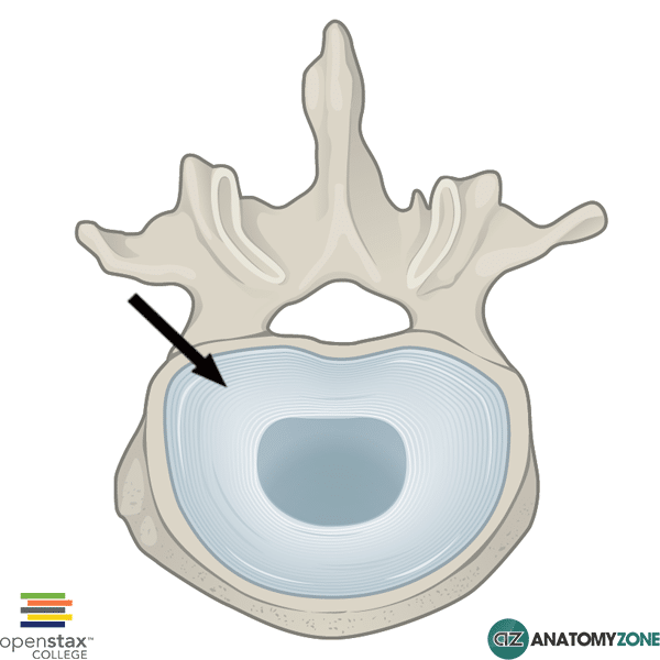
The structure indicated is the anulus fibrosus of the intervertebral disc.
Intervertebral discs are found between adjacent vertebral bodies in the spinal column. Intervertebral discs are one of two major joint types found in the vertebrae, the other type being synovial joints between the articular processes.
The symphyses between the vertebral bodies consists of hyaline cartilage which lines the vertebral body, and an intervertebral disc which is sandwiched between the two adjacent intervertebral bodies to form this fibrocartilaginous joint.
The intervertebral disc consist of
The anulus fibrosus forms the outer fibrous portion, which envelops the inner gel-like nucleus pulposus. The anulus fibrosus consists of laminae of fibrous tissue and fibrocartilage – the organisation of this tough tissue into lamellae allow the intervertebral discs to sustain heavy compressive loads.
Disc prolapse occurs when the inner gel-like nucleus pulposus is forced out of the anulus fibrosis, thereby exerting pressure on local nerves, or the spinal cord.
Learn more about the anatomy of the vertebrae in this tutorial!