Quantcast
Channel: AnatomyZone
Viewing all 279 articles
Browse latest View live

Superior Oblique Muscle
$
0
0

Superior Oblique Muscle

Superior Oblique Muscle

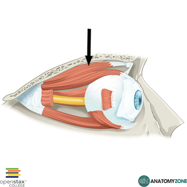
The structure indicated is the superior oblique muscle.

The superior oblique muscle is one of the extra-ocular muscles. The extra-ocular muscles include the medial, lateral, superior and inferior recti muscles, and the superior and inferior oblique muscles.

Origin: Annulus of Zinn

Insertion: Outer posterior quadrant of the eyeball

Innervation: Trochlear nerve

Action: Moves the eyeball down and out (depression, abduction, medial rotation).

The superior oblique muscle inserts onto the eyeball via a long tendon which loops around a pulley (the trochlea of the superior oblique) on the medial aspect of the orbital roof, just lateral to the insertion point of the superior rectus muscle. The superior oblique muscle is innervated by the fourth cranial nerve, the trochlear nerve – this cranial nerve has the longest intracranial course and is susceptible to injury.

Fourth nerve palsy causes weakness or paralysis of the superior oblique muscle, causing vertical diplopia (double vision) that is worse on downward lateral gaze. Patients with fourth nerve palsy classically tilt their head away from the affected side to compensate for the diplopia.
Cortical Homunculus

$
0
0

Cortical Homunculus

Sensory Homonculus

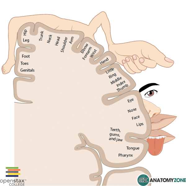
The diagram illustrates the cortical homunculus.

The cortical homunculus visually portrays the anatomical divisions of the primary motor cortex and the primary somatosensory cortex. The word “homunculus” comes from the Latin word which translates as “little man”.

It represents how our body is perceived within the brain and how the neurons are distributed in these areas in order to create this perception. What immediately stands out is that the size of a body area is not proportional to its representation in the cortical homunculus – certain areas such as the hands, lips and face occupy disproportionately large areas of the cortex, due to the importance of fine motor skills and and the extensive innervation of these areas.

Knowledge of the cortical homunculus can help to explain the pattern of motor and sensory symptoms in patients who have suffered a stroke. The middle cerebral artery, broadly speaking supplies the lateral cerebral cortex. Looking at the cortical homunculus you can see that the lateral parts of the cerebrum in the motor and sensory cortices represent the upper limbs and facial structures. Occlusion of the middle cerebral artery therefore, can lead to contralateral paralysis/paraparesis (weakness) and/or sensory loss of the contralateral face and arm.

The anterior cerebral artery, on the other hand, supplies the medial aspect of the cerebral hemispheres. You can therefore infer from the cortical homunculus, that occlusion of the anterior cerebral artery can lead to sensory and motor deficits in the leg and foot.

Learn more about the anatomy of the brain in this tutorial, and learn about the blood supply to the brain in this tutorial.

Hypoglossal Nerve

$
0
0

Hypoglossal Nerve

Hypoglossal Nerve

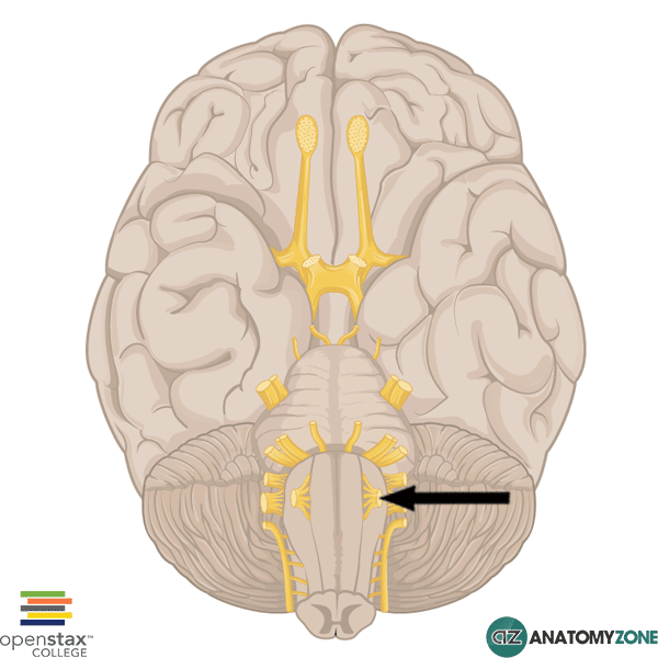
The structure indicated is the hypoglossal nerve (cranial nerve XII).

The hypoglossal nerve carries general somatic efferent nerve fibres and is responsible for motor innervation to the extrinsic and intrinsic muscles of the tongue. It arises from the hypoglossal nucleus located in the caudal brain stem and emerges from the preolivary sulcus in the ventromedial aspect of the medulla oblongata from several rootlets. The preolivary sulcus separates the olive from the medullary pyramids. The hypoglossal nerve then passes through the subarachnoid space and exits the skull through the hypoglossal canal.

The extrinsic muscles of the tongue are as follows:

  • Genioglossus
  • Hyoglossus
  • Styloglossus
  • Palatoglossus

Damage to the hypoglossal nerve causes weakness of the tongue muscles resulting in deviation of the tongue towards the side of the lesion.

Learn all about the cranial nerves in this anatomy tutorial.

Learn all about the extrinsic muscles of the tongue in this anatomy tutorial.

Orbicularis Oris Muscle

$
0
0

Orbicularis Oris

Orbicularis Oris

The muscle indicated is the orbicularis oris muscle.

The orbicularis oris muscle is one of the muscles of facial expression belonging to the oral group. The muscles of facial expression can be organised into functional groups to provide a more structured approach to understanding the muscles. The functional groups include the following:

The oral group of muscles includes muscles that are responsible for moving the lips and the cheek. There are 11 muscles included in this functional group: orbicularis oris, depressor anguli oris, depressor labi inferioris, mentalis, risorius, buccinator, zygomaticus major, zygomaticus minor,  levator labii superioris, levator labii superioris alaeque nasi, levator anguli oris.

The muscle fibres of the orbicularis oris form a circular ring around the mouth. Contraction of the orbicularis oris results in closing of the lips and narrowing of the mouth – “pursing of the lips”. It receives innervation from the buccal branch of the facial nerve (cranial nerve VII).

To learn more about the anatomy of the muscles of facial expression, watch this tutorial.

Tunica Vaginalis

$
0
0

Tunica Vaginalis

Tunica Vaginalis

The structure indicated is the tunica vaginalis.

The tunica vaginalis forms the serous covering of the testis, originating in embryological life from the processus vaginalis of the peritoneum (an embryonic outpouching of the peritoneum). In fetal life, the processus vaginalis precedes the descent of the testis into the scrotum from the abdomen and then closes off. The remaining part then becomes known as the tunica vaginalis which adheres to the surface of the testis and reflects back onto the internal aspect of the scrotum. It therefore consists of a visceral portion and a parietal portion. The visceral portion adhering to the testis and the parietal potion adhering to the internal scrotal wall.

If the processus vaginalis fails to close off, peritoneal fluid can therefore travel into the scrotum leading to the formation of a hydrocele.

Learn more about the anatomy of the testis and male reproductive system in this tutorial.

Suspensory Ligament of Breast (Cooper’s Ligaments)

$
0
0

Suspensory Ligament of Breast (Cooper’s Ligaments)

Suspensory Ligament of Breast (Cooper's Ligaments)

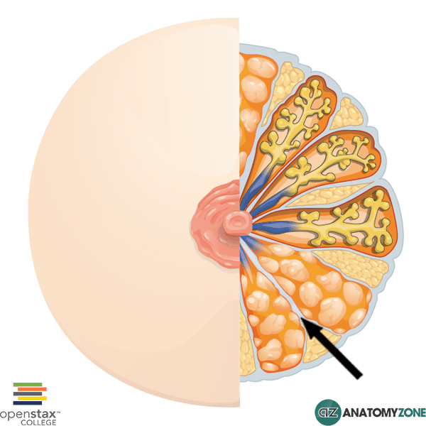
The structure indicated is the suspensory ligament of the breast, also known as Cooper’s ligaments.

These ligaments are formed from the connective tissue stroma which surround the ducts and lobules of the mammary gland – in certain areas the connective tissue condenses forming ligaments which support the breast, extending from the dermis of the overlying skin to the deep fascia. These suspensory ligaments support the breast on the chest wall.

The suspensory ligaments of Cooper give rise to the dimpling appearance (peau d’orange) that is seen following development of inflammatory carcinoma, as the lymphatic ducts become blocked and the skin remains tethered by the ligaments.

Iliacus Muscle

$
0
0

Iliacus Muscle

Iliacus Muscle

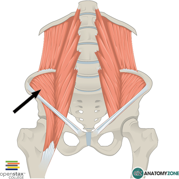
The muscle indicated is the iliacus muscle.

The iliacus originates in the iliac fossa on the inside of the pelvic bone. The iliacus combines with the psoas major to enter the anterior compartment of the thigh and insert via a common tendon on the lesser trochanter of the femur. The iliacus and psoas are thus collectively referred to as the iliopsoas muscle – they act synergistically to flex the thigh at the hip joint, as well as acting to cause lateral rotation of the thigh. The iliopsoas muscles are the strongest hip flexors.

Origin: Iliac fossa

Insertion: Base of the lesser trochanter of the femur

Innervation: Femoral nerve

Action: Flexion of the thigh at the hip joint. Lateral rotation of the thigh.

Learn more about the iliacus muscle and the muscles of the anterior compartment of the thigh in this anatomy tutorial.

Precentral Gyrus

$
0
0

Precentral Gyrus

Precentral Gyrus

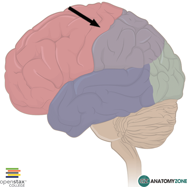
The structure indicated is the precentral gyrus.

A gyrus is a ridge on the cerebral cortex, and is usually surrounded by grooves, known as sulci. Fissures are larger than sulci and divide the brain into lobes, as well as dividing the brain into right and left hemispheres.

The sulci anterior to the precentral gyrus is known as the precentral sulcus. This sulcus lies parallel to the central sulcus (also referred to as the fissure of Rolando/Rolandic fissure). The central sulcus separates the parietal lobe from the frontal lobe. The precentral gyrus contains the primary somatomotor cortex.

Learn more about the precentral gyrus in this anatomy tutorial on the basic parts of the brain.


Major alar cartilage

$
0
0

Major alar cartilage

Major alar cartilage

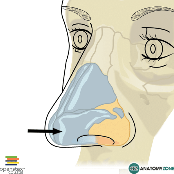
The structure indicated is the major alar cartilage (greater alar cartilage).

The lateral walls of the external nose are comprised from three cartilages:

  1. Lateral process of septal cartilage
  2. Major alar cartilage
  3. Minor alar cartilage

The major alar cartilage is located directly below the lateral process of the septal cartilage. It is structured such that it is bent in on itself to form both the medial and lateral walls of the nose. The crus laterale forms the lateral wall, whereas the crus mediale forms the medial wall.

Minor Calyx of Kidney

$
0
0

Minor Calyx of Kidney

Minor Calyx of Kidney

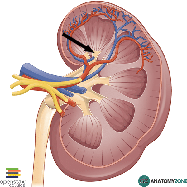
The structure indicated is the minor calyx of the kidney.

The kidney consists of an outer cortex and an inner medulla. The cortex projects into the kidney dividing the medulla into triangular shaped renal pyramids, whose apices are surrounded by a minor calyx (calices = plural). The minor calices join to form a major calyx, which in turn unite to form the renal pelvis which exit the kidney to form the ureters. The calyx system of the kidney serves to drain urine. The smooth muscle walls of the calices are capable of peristalsis, which helps to propel urine through the kidneys, out through the renal pelvis and into the bladder via the ureters.

Learn all about the anatomy of the internal structure of the kidney in this video tutorial.

Iliocostalis Lumborum

$
0
0

Iliocostalis Lumborum

Iliocostalis Lumborum

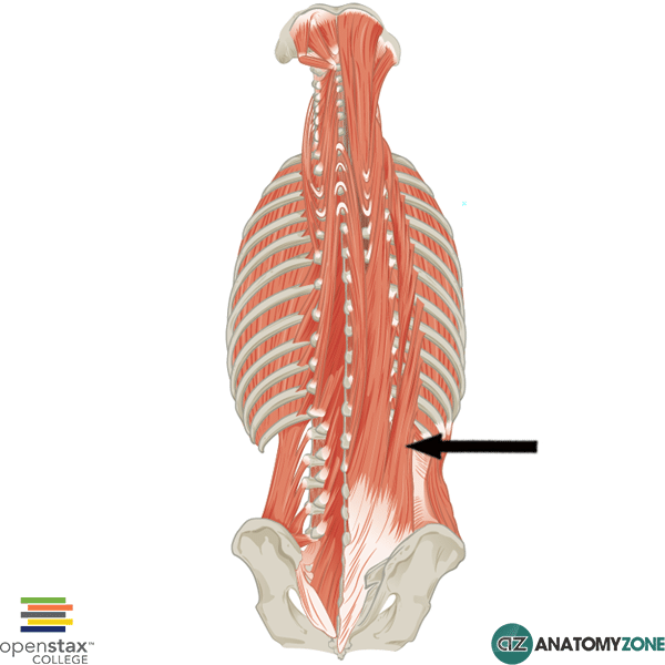
The muscle indicated is the iliocostalis lumborum muscle, one of the erector spinae group of back muscles.

The back muscles can be divided into three groups of muscles:

  • Superficial
  • Intermediate
  • Deep group

The erector spinae muscles belong to the deep (intrinsic) group of back muscles and are the largest group of intrinsic back muscles. There are three main erector spinae muscles, from lateral to medial:

  • Iliocostalis
  • Longissimus
  • Spinalis

Each of these three muscles is further subdivided into separate parts:

  • Lumborum
  • Thoracis
  • Cervicis
  • Capitis

Origin: sacrum, iliac crest, spinous processes of lumbar vertebrae and thoracic vertebrae T11 and T12

Insertion: Inferior borders of lower 6/7 ribs

Innervation: Posterior branch of spinal nerve

Action: Unilateral contraction – flexes head to same side. Bilateral contraction – extension of vertebral column

Learn all about the anatomy of the superficial back muscles and the intermediate and deep back muscles.

Superior Sagittal Sinus

$
0
0

Superior Sagittal Sinus

Superior Sagittal Sinus

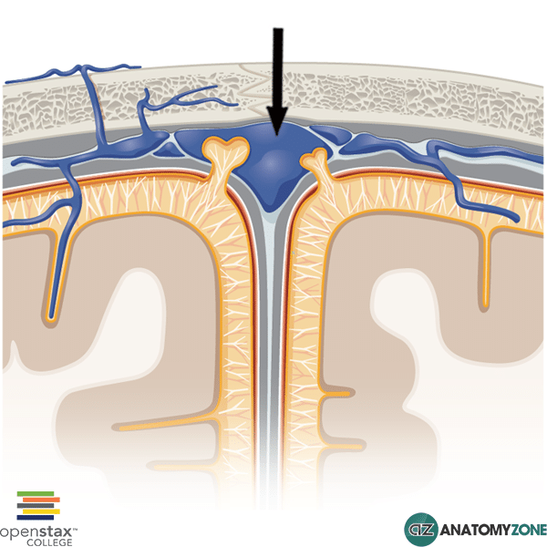
The structure indicated is the superior sagittal sinus.

The brain is drained by a series of veins and venous channels which drain into large dural venous sinuses, which in turn ultimately drain to the internal jugular veins. The dural venous sinuses are lined by endothelium and located between the layers of the dura mater in the brain. The venous sinuses are different to other blood vessels as they do not have the same set of layers which form their walls, and do not contain valves, like veins.

The venous sinuses receive blood from veins which drain the brainstem, the cerebrum and the cerebellum, as well as diploic and emissary veins. Diploic veins run from inside the compact bone of the cranium into the venous sinuses, whereas emissary veins run from outside the cranium through the compact bone and into the venous sinuses.

The superior sagittal sinus is located on the superior border of the falx cerebri and receives blood from superior cerebral, diploic, and emissary veins, as well as receiving CSF via arachnoid granulations. It drains blood from lateral and anterior parts of the cerebrum into the confluence of sinuses.

The superior sagittal sinus beings at the foramen cecum and drains into the confluence of sinuses near the internal occipital protuberance.

Pseudostratified Columnar Epithelium

$
0
0

Pseudostratified Columnar Epithelium

Pseudostratified Columnar Epithelium

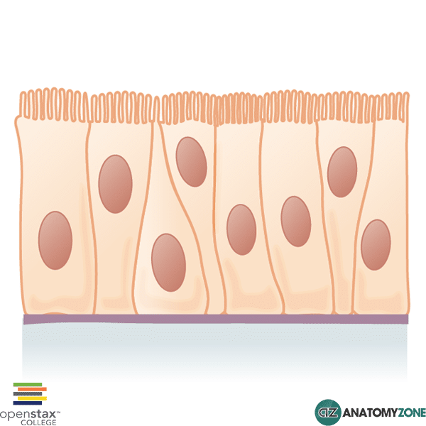
This diagram represents pseudostratified columnar epithelium.

This is a special type of simple epithelium called pseudostratified epithelium as it resembles stratified epithelium due to the positioning of the cellular nuclei, but is comprised of only a single layer of cells. Pseudostratified epithelium is most commonly found in the form of columnar shaped epithelium, but can also be formed from squamous or cuboidal epithelia.

Ciliated pseudostratified columnar epithelia is found lining the trachea and upper respiratory tract.

Non-ciliated pseudostratified columnar epithelia is found in the membranous part of the vas deferens and also lining the epididymis of the male reproductive system.

Eponychium

$
0
0

Eponychium

Eponychium

The structure indicated is the eponychium of the fingernail.

The eponychium is a thickened layer of skin which surrounds the nails of the fingers and toes. It serves to protect the area between the nail and the epidermis from infection. The eponychium is often confused with the cuticle. The cuticle is the visible fold of dead skin that forms a semi-circular shape on the proximal nail plate, and it is this layer of dead skin that is removed during a manicure. The eponychium is the living fold of skin which produces the cuticle.

Epiphyseal Line

$
0
0

Epiphyseal Line

Epiphyseal Line

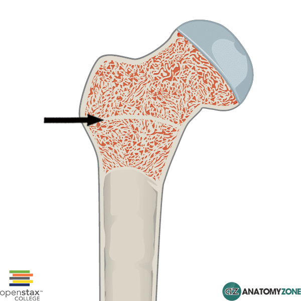
The structure indicated is the epiphyseal line.

The epiphyseal plate is a plate of hyaline cartilage found in children and adolescents, located in the metaphysis at the ends of each long bone. Long bones consist of a diaphysis, metaphysis and epiphysis. The diaphysis is the main long section of the bone, the epiphysis is the rounded end of the long bone, and the metaphysis is the section of bone between the diaphysis and metaphysis.  In adults, the epiphyseal plate is replaced with the epiphyseal line and marks the point of union were the epiphysis meets the diaphysis.

The epiphyseal plates permit growth and lengthening of the bone, as the cartilage reproduces and ossifies. The epiphyseal plate has a zonal arrangement, with cartilaginous proliferation occurring closer to the epiphysis, and ossification occurring closer to the diaphysis. The zonal arrangement of the epiphyseal plate, from epiphysis to diaphysis, is as follows:

  • Zone of reserve
  • Zone of proliferation
  • Zone of maturation and hypertrophy
  • Zone of calcification
  • Zone of ossification

Stapes (ossicle of ear)

$
0
0

Stapes (ossicle of ear)

Stapes (ossicle of ear)

The ossicles of the ear are tiny little bones that are located in the middle ear. They are the smallest bone in the human body (the word itself actually means “little bone”) and their purpose is to transmit sound vibrations from the eardrum (tympanic membrane), to the complex inner ear structures, ultimately leading to the perception of sound.

There are a series of three ossicles in the ear:

  • malleus
  • incus
  • stapes.

The malleus is attached to the tympanic membrane and articulates with the incus, which in turn articulates with the stapes. The stapes, attaches to the membrane of the oval window connecting the middle ear to the inner ear. Through this series of bones, the vibrations are transmitted from the tympanic membrane to the inner ear.

The stapes is a stirrup-shaped bone (it gets its name from the Latin Stapes which means stirrup), and is the smallest and lightest bone in the human body.

The stapes consists of the following components:

  • Base
  • Anterior crus
  • Posterior crus
  • Head
  • Neck

The base of the stapes is connected to the oval window via the annular ligament. The head of the stapes articulates with the incus via the incudostapedial joint.

Spinothalamic Tract

$
0
0

Spinothalamic Tract

Spinothalamic Tract

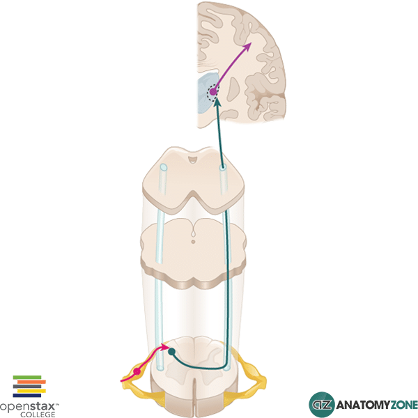
This diagram illustrates the spinothalamic tract (also known as the anterolateral system/ventrolateral system).

The spinothalamic tract is one of the ascending sensory tracts which conveys sensory information from the periphery to the central processing systems. There are three main ascending (afferent) pathways which convey sensory information:

  1. Dorsal column medial lemniscus system
  2. Spinocerebellar tracts
  3. Spinothalamic tracts (anterolateral system)

The spinothalamic tract is responsible for conveying the modalities of crude touch, pressure, pain and temperature. The tract is comprised of a three neurone sequence, which terminates in the somatosensory cortex of the postcentral gyrus:

  • First order neurone: information carried from peripheral sensory receptors to spinal cord via pseudounipolar neurones. Axons ascend 1-2 vertebral levels and synapse with second order neurones in the substantia gelatinosa.
  • Second order neurones: immediately decussate (cross over to the other side) via the anterior white commissure, and ascend to the medulla of the brainstem and then to the thalamus where they synapse with third order neurones.
  • Third order neurones: project to the primary somatosensory cortex

The anterior spinothalamic tract conveys crude touch.

The lateral spinothalamic tract conveys pain and temperature.

Ciliary Body

$
0
0

Ciliary Body

Ciliary Body

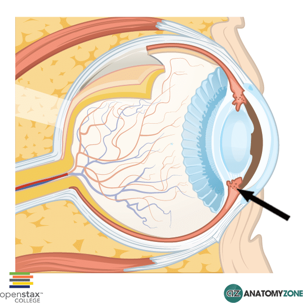
The structure indicated is the ciliary body.

The eyeball consists of a fibrous outer layer, a vascular layer, and an inner layer.

The vascular layer of the eyeball consists of:

  • Choroid
  • Ciliary body
  • Iris

The ciliary body is triangular in cross-section and encircles the eyeball. It has two components:

  • Ciliary muscle
  • Ciliary processes

The ciliary body has three main functions:

  • Accommodation of the lens
  • Production and resorption of aqueous humor
  • Maintenance of lens zonules

Fungiform Papilla

$
0
0

Fungiform Papilla

Fungiform Papilla

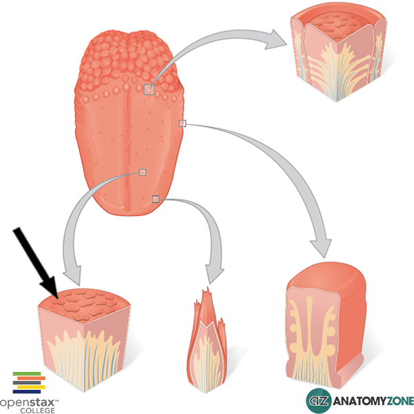
The structure indicated represents a fungiform papillae, one of the four types of lingual papillae found on the surface of the tongue.

There are four types of lingual papillae found on the tongue:

  • Circumvallate papillae (top right)
  • Fungiform papillae (bottom left)
  • Filiform papillae (bottom middle)
  • Foliate papillae (bottom right)

The only papillae not associated with taste buds are the filiform papillae. Fungiform papillae are mushroom shaped and contain taste buds on their upper surface allowing distinction of all five modalities of taste:

  • Bitter
  • Sweet
  • Sour
  • Salty
  • Umami

Splenic Artery

$
0
0

Splenic Artery

Splenic Artery

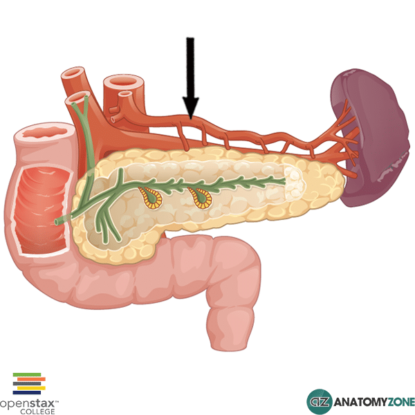
The structure indicated is the splenic artery.

The splenic artery the largest of three arteries arising for the celiac trunk:

  • Left gastric artery
  • Common hepatic
  • Splenic artery

The celiac artery is responsible for supplying the foregut structures, arising from the abdominal aorta just below the point at which it emerges through the aortic hiatus, at the level of vertebra L1.

The splenic artery takes a tortuous course along the superior aspect of the pancreas to enter the hilum of the spleen. Along its course, it gives off several branches supplying the neck, body and tail of the pancreas.

The splenic artery gives off the following arteries:

  • Short gastric arteries
  • Left gastro-omental artery (left gastroepiploic)
  • Pancreatic branches

The short gastric arteries supply the fundus of the stomach. The left gastro-omental artery passes medially along the greater curvature of the stomach to anastomose with the right gastro-omental artery (a branch of the gastroduodenal artery, which arises from the common hepatic artery).

Learn about the anatomy of the foregut blood supply and the celiac trunk in this video tutorial.

Viewing all 279 articles
Browse latest View live