Quantcast
Channel: AnatomyZone
Viewing all 279 articles
Browse latest View live

Dorsal Interossei

$
0
0

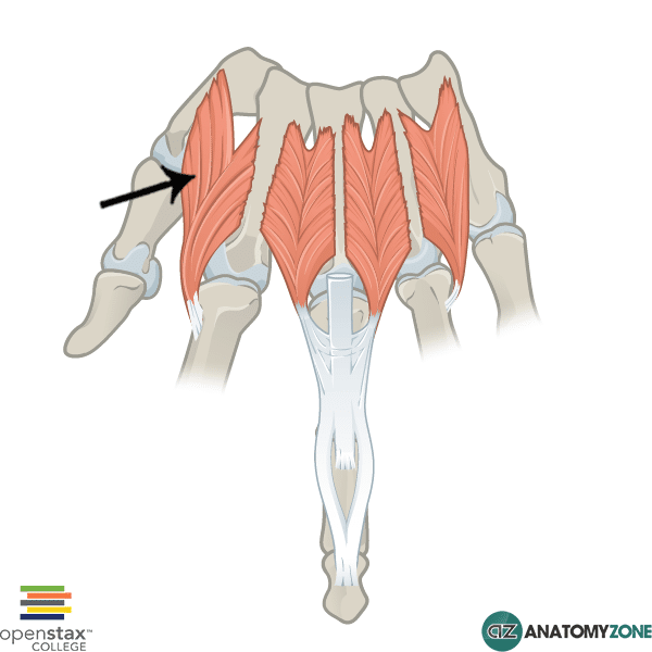
Dorsal Interossei

First dorsal interosseus muscle

The structure indicated is the first dorsal interosseous muscle of the hand.

The dorsal interossei belong to the intrinsic group of muscles which act on the hand. The intrinsic muscles include the following muscles/groups:

  • Thenar group (act on thumb)
  • Hypothenar group (act on little finger)
  • Adductor pollicis
  • Lumbricals
  • Interossesus muscles (dorsal and palmar)
  • Palmaris brevis

All the intrinsic muscles of the hand, except the thenar muscles and the lateral two lumbrical muscles are innervated by the deep branch of the ulnar nerve. The thenar muscles and the lateral two lumbrical muscles are innervated by the median nerve.  A useful mnemonic for remembering this is MEATLOAF. “MEAT” refers to the Median nerve, and LOAF refers to the muscles which it innervates: Lateral two lumbricals, Opponens pollicis, Abductor pollicis brevis, Flexor pollicis brevis.

The interosseous muscles, as the name suggests are located between (inter-) the metacarpal bones (-osseus). There are 4 bipennate dorsal interossei located between the adjacent shafts of the metacarpal bones and insert onto the bases of the proximal phalanges and the extensor hoods. The dorsal interossei serve to abduct the fingers away from the central axis of the middle finger. The first interosseous muscle inserts onto the base laterally on the index finger.

The palmar interossei in contrast are unipennate in structure and function to adduct the fingers towards the central axis of the middle finger. A useful mnemonic for remembering this function is PAD and DAB: Palmar interossei AD-duct, Dorsal interossei AB-duct.

Learn more about the anatomy of the hand muscles in this tutorial.

Pisiform Bone

$
0
0

Pisiform Bone

Pisiform Bone

The structure indicated is the pisiform bone of the hand.

There are three groups of bones in the hand:

  1. Carpal bones (8 in total)
  2. Metacarpal bones
  3. Phalanges

The carpal bones are separated into two rows:

  1. Proximal row
  2. Distal row

The pisiform bone is one of four bones in the proximal row of carpals (scaphoid, lunate, triquetral, pisiform). The pisiform bone is a tiny sesamoid bone located in the tendon of the flexor carpi ulnaris muscle. It articulates with the palmar surface of the triquetral bone.

Learn more about the anatomy of the bones of the hand in this tutorial.

Episode 7 – Cardiac Muscle

$
0
0
Learn all about the physiology of the cardiac muscle in this brand new physiology tutorial.

Quadratus Femoris

$
0
0

Quadratus Femoris

Quadratus Femoris

The structure indicated is the quadratus femoris muscle.

The quadratus femoris muscle is one of the muscles of the gluteal region. The muscles in the gluteal region are divided into a superficial and deep group. There are four muscles of the superficial group:

  1. Gluteus maximus
  2. Gluteus medius
  3. Gluteus minimus
  4. Tensor fasciae latae

There are 5 muscles in the deep group:

  1. Gemellus superior
  2. Gemellus inferior
  3. Quadratus femoris
  4. Piriformis
  5. Obturator internus

The deep muscles of the thigh are much smaller than the superficial group of muscles and generally serve to laterally rotate the femur at the hip joint.

The quadratus femoris muscle is the most inferiorly located muscle in the deep gluteal region of muscles and is named according to its approximately rectangular shape.

Origin: Lateral margin of obturator ring (above ischial tuberosity)

Insertion: Quadrate tubercle and intertrochanteric crest of proximal posterior femur

Action: Lateral rotation of hip. Assists with adduction of hip

Innervation: Nerve to quadratus femoris

Learn more about the anatomy of the thigh and gluteal muscles in this tutorial.

Cervical Plexus

$
0
0

This video was made in collaboration with TeachMeAnatomy.info, check out the related article to consolidation your learning on this topic.

Axillary Nerve

$
0
0

This video was made in collaboration with TeachMeAnatomy.info, check out the related article to consolidation your learning on this topic.

Ulnar Nerve

$
0
0

This video was made in collaboration with TeachMeAnatomy.info, check out the related article to consolidation your learning on this topic.

Median Nerve

$
0
0

This video was made in collaboration with TeachMeAnatomy.info, check out the related article to consolidation your learning on this topic.


Radial Nerve

$
0
0

This video was made in collaboration with TeachMeAnatomy.info, check out the related article to consolidation your learning on this topic.


In this video the following structures are discussed:

  • Radial nerve
  • Posterior cord
  • Posterior cutaneous nerve of arm
  • Posterior cutaneous nerve of forearm
  • Inferior lateral cutaneous nerve of arm
  • Superficial and deep branches of radial nerve
  • Triangular interval

Angiogenesis

$
0
0

Angiogenesis

Illustration of the progression of angiogenesis.

Angiogenesis is the physiological process of new blood vessel growth from PRE-EXISTING vessels. This is a different process from that of NEW blood vessel growth, which is called vasculogenesis.

Angiogenesis is a fascinating process that is involved in both normal physiology (wound healing and formation of granulation tissue) and also in the pathological process of tumour transformation. Tumours require a rich blood supply in order to grow and continue rapidly dividing. To enable further growth angiogenesis is critical, and is therefore a key step in the transformation of a benign tumour into a malignant one.

For this reason, inhibition of angiogenesis is a key target of anti-tumour cancer therapies.

Image credit: Dr Jon Heras

Musculocutaneous Nerve

$
0
0

This video was made in collaboration with TeachMeAnatomy.info, check out the related article to consolidation your learning on this topic.


In this video the following structures are discussed:

  • Musculocutaneous nerve
  • Lateral cutaneous nerve of forearm (lateral antebrachial cutaneous nerve)

Sacral Plexus

$
0
0

This video was made in collaboration with TeachMeAnatomy.info, check out the related article to consolidation your learning on this topic.


In this video the following structures are discussed:

  • Lumbar plexus
  • Superior gluteal nerve
  • Inferior gluteal nerve
  • Sciatic nerve
  • Posterior femoral cutaneous nerve
  • Pudendal nerve
  • Nerve to quadratus femoris
  • Nerve to obturator internus
  • Perforating cutaneous nerve
  • Nerve to piriformis

Orbit Bones – Eye

$
0
0

This video was made in collaboration with TeachMeAnatomy.info, check out the related article to consolidation your learning on this topic.

In this video the following are discussed:

  • Orbital Rim
  • Boundaries of orbit
  • Bones of orbit
  • Contents of orbit

Extraocular Muscles – Eye

$
0
0

This video was made in collaboration with TeachMeAnatomy.info, check out the related article to consolidation your learning on this topic.

In this video the following are discussed:

  • Superior rectus
  • Inferior rectus
  • Medial rectus
  • Lateral rectus
  • Superior oblique
  • Inferior oblique
  • Movements of eye: intorsion, extorsion, abduction, adduction, elevation, depression

Eyeball

$
0
0

This video was made in collaboration with TeachMeAnatomy.info, check out the related article to consolidation your learning on this topic.

In this video the following structures are discussed:
- sclera
- cornea
- choroid
- iris
- pupil
- suspensory ligament
- lens
- ciliary muscle
- ciliary processes
- ciliary body
- retina
- anterior chamber
- posterior chamber
- scleral venous sinus
- vitreous humour
- aqueous humour
- optic disc
- retinal branch arteries
- macula lutea
- fovea centralis
- zonular fibres


Eyeball – Blood Supply

$
0
0

This video was made in collaboration with TeachMeAnatomy.info, check out the related article to consolidation your learning on this topic.

In this video the following structures are discussed:

  • Internal carotid artery
  • Ophthalmic artery
  • Short posterior ciliary arteries
  • Long posterior ciliary arteries
  • Anterior ciliary arteries
  • Central retinal artery
  • Optic disc
  • Cavernous sinus
  • Superior ophthalmic vein
  • Inferior ophthalmic vein
  • Vorticose veins

Ulnar Nerve

$
0
0

This video was made in collaboration with TeachMeAnatomy.info, check out the related article to consolidation your learning on this topic.

Median Nerve

$
0
0

This video was made in collaboration with TeachMeAnatomy.info, check out the related article to consolidation your learning on this topic.

Radial Nerve

$
0
0

This video was made in collaboration with TeachMeAnatomy.info, check out the related article to consolidation your learning on this topic.


In this video the following structures are discussed:

  • Radial nerve
  • Posterior cord
  • Posterior cutaneous nerve of arm
  • Posterior cutaneous nerve of forearm
  • Inferior lateral cutaneous nerve of arm
  • Superficial and deep branches of radial nerve
  • Triangular interval

Angiogenesis

$
0
0

Angiogenesis

Angiogenesis

Illustration of the progression of angiogenesis.

Angiogenesis is the physiological process of new blood vessel growth from PRE-EXISTING vessels. This is a different process from that of NEW blood vessel growth, which is called vasculogenesis.

Angiogenesis is a fascinating process that is involved in both normal physiology (wound healing and formation of granulation tissue) and also in the pathological process of tumour transformation. Tumours require a rich blood supply in order to grow and continue rapidly dividing. To enable further growth angiogenesis is critical, and is therefore a key step in the transformation of a benign tumour into a malignant one.

For this reason, inhibition of angiogenesis is a key target of anti-tumour cancer therapies.

Image credit: Dr Jon Heras

Viewing all 279 articles
Browse latest View live