Quantcast
Channel: AnatomyZone
Viewing all 279 articles
Browse latest View live

Acromioclavicular Joint

$
0
0

Acromioclavicular Joint

Acromioclavicular Joint

The structure indicated is the acromioclavicular joint.

The shoulder joint complex consists of the three joints:

  1. Glenohumeral joint
  2. Acromioclavicular joint
  3. Sternoclavicular joint

The acromioclavicular joint is formed between the acromion of the scapula and the clavicle. It is a small synovial joint that is stabilised by the following ligaments:

  • Acromioclavicular ligament (consists of superior and inferior parts)
  • Coracoclavicular ligaments

The coracoclavicular ligaments consist of two parts:

  • Trapezoid ligament
  • Conoid ligament

Learn more about the shoulder joint in this anatomy tutorial.


Internal Carotid Artery

$
0
0

Internal Carotid Artery

Internal Carotid Artery

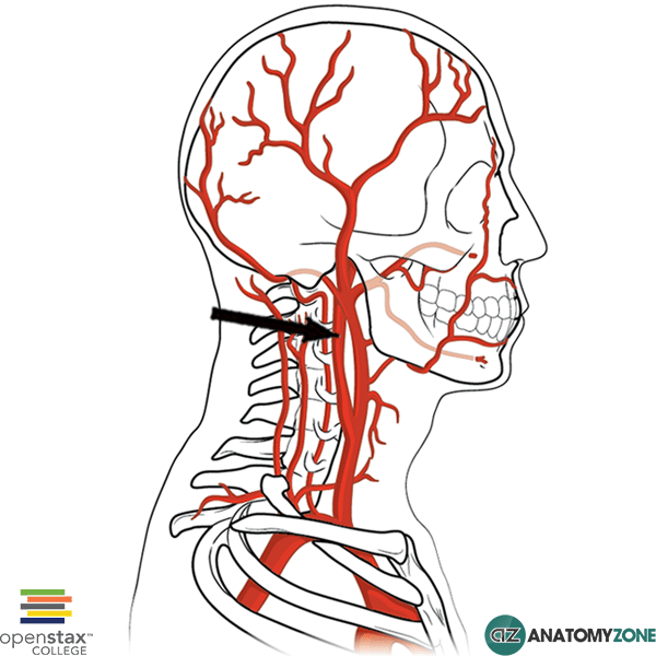
The structure indicated is the internal carotid artery.

The internal carotid arteries arise from the common carotid arteries.

The brachiocephalic artery arises from the arch of the aorta and divides into the right subclavian artery and the right common carotid artery.

There is no left brachiocephalic artery however, and the left common carotid arises directly from the aortic arch. There are three branches which come off the aortic arch:

  1. Brachiocephalic artery
  2. Left common carotid
  3. Left subclavian

The common carotid arteries then divide into the external carotid and internal carotid arteries. The internal carotid artery ascends to supply the brain, joining the circle of Willis. The external carotid artery supplies the structures of the head and face.

The internal carotid arteries consists of 7 segments:

  1. Cervical segment (C1)
  2. Petrous segment (C2)
  3. Lacerum segment (C3)
  4. Cavernous segment (C4)
  5. Clinoid segment (C5)
  6. Ophthalmic segment (C6)
  7. Communicating segment (C7)

Episode 4 – Muscle Contraction

$
0
0
Learn all about the physiology of muscle contraction in this brand new physiology tutorial.

Episode 5 – Muscle Metabolism

$
0
0
Learn all about the physiology of muscle metabolism in this brand new physiology tutorial.

Coracoclavicular Ligament

$
0
0

Coracoclavicular Ligament

Coracoclavicular Ligament

The structure indicated is the coracoclavicular ligament

The coracoclavicular ligament is one of the ligaments which stabilises the acromioclavicular joint. The acromioclavicular joint is formed between the acromion of the scapula and the clavicle. It is a small synovial joint that is stabilised by the following ligaments:

  • Acromioclavicular ligament (consists of superior and inferior parts)
  • Coracoclavicular ligaments

The coracoclavicular ligaments consist of two parts:

  • Trapezoid ligament
  • Conoid ligament

The coracoclavicular ligament, as the name suggests connects the clavicle to the coracoid process of the scapula.

Learn more about the anatomy of the shoulder joint in this tutorial.

Posterior Inferior Tibiofibular Ligament

$
0
0

Posterior Inferior Tibiofibular Ligament

Posterior Inferior Tibiofibular Ligament

The structure indicated is the posterior inferior tibiofibular ligament.

The distal ends of the tibia and the fibula are held together by the inferior portion of the interosseous membrane. The interosseous membrane is the membrane which joins the tibia to the fibula along its entire length, separating the leg into anterior and posterior compartments. There are two apertures within the interosseous membrane proximally and distally which allow vessels to pass from one compartment to the other.

In addition to the attachment of the interosseous membrane inferiorly between the distal tibia and fibula, this inferior articulation is reinforced by the tibiofibular ligaments anteriorly and posteriorly.

Learn more about the anatomy of the tibia and fibula in this tutorial.

Ischiocavernosus

$
0
0

Ischiocavernosus

Ischiocavernosus
Ischiocavernosus

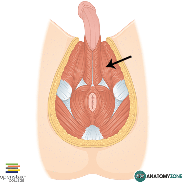
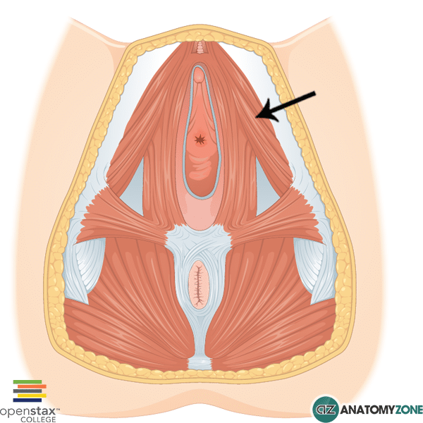
The structure indicated is the ischiocavernosus muscle.

The ischiocavernosus muscle is one of three pairs of muscles in the superficial perineal pouch:

  1. Ischiocavernosus
  2. Bulbospongiosus
  3. Superficial transverse perineal muscles

It originates from the inner aspect of the ischiopubic ramus and attaches on either side to the crura of the penis and clitoris.

Origin: Ischiopubic ramus – internal aspect

Insertion: Crus of penis and clitoris

Action: Stabilise erect penis

Innervation: Pudendal nerve

Learn more about the pelvic floor in this tutorial

Transverse Foramen

$
0
0

Transverse Foramen

Transverse Foramen

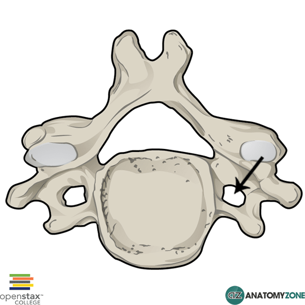
The structure indicated is the transverse foramen of a cervical vertebra.

There are 33 vertebrae which make up the spinal column:

  • 7 cervical
  • 12 thoracic
  • 5 lumbar
  • 5 fused sacral vertebrae
  • 3-4 fused coccygeal vertebrae

The typical vertebra consists of the following features:

  • Vertebral body
  • Vertebral arch (neural arch) consisting of lamina and pedicle
  • Spinous process
  • Transverse process
  • Vertebral foramen (collectively form vertebral canal)
  • Superior and inferior articular processes
  • Superior and inferior vertebral notches

Transverse foramina are only present in the cervical vertebrae. These foramina allow the passage of the vertebral artery and vein.

The vertebral arteries arise from the first part of the subclavian artery. The vertebral and internal carotid arteries provide the arterial supply to the brain, forming the Circle of Willis at the base of the brain.

After the vertebral artery is given off from the subclavian artery, it passes through the transverse foramina of cervical vertebrae C1 to C6. It then enters the skull via the foramen magnum.

Learn more about the anatomy of the vertebrae in this tutorial.


Gluteus Minimus

$
0
0

Gluteus Minimus

Gluteus Minimus

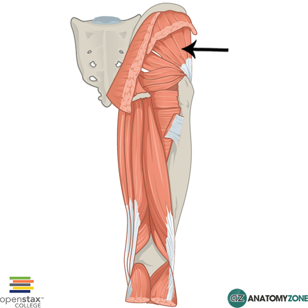
The structure indicated is the gluteus minimus.

The gluteus minimus muscle is one of the muscles of the gluteal region. The muscles in the gluteal region are divided into a superficial and deep group. There are four muscles of the superficial group:

  1. Gluteus maximus
  2. Gluteus medius
  3. Gluteus minimus
  4. Tensor fasciae latae

There are 5 muscles in the deep group:

  1. Gemellus superior
  2. Gemellus inferior
  3. Quadratus femoris
  4. Piriformis
  5. Obturator internus

Origin: Dorsal ilium between inferior and anterior gluteal lines and edge of greater sciatic notch.

Insertion: lateral and superior surfaces of greater trochanter

Action: Hip abduction, hip medial rotation (anterior fibres), hip lateral rotation (posterior fibres)

Innervation: Superior gluteal nerve

Learn more about the anatomy of the gluteal muscles in this tutorial.

Superior Articular Process

$
0
0

Superior Articular Process

Superior Articular Process

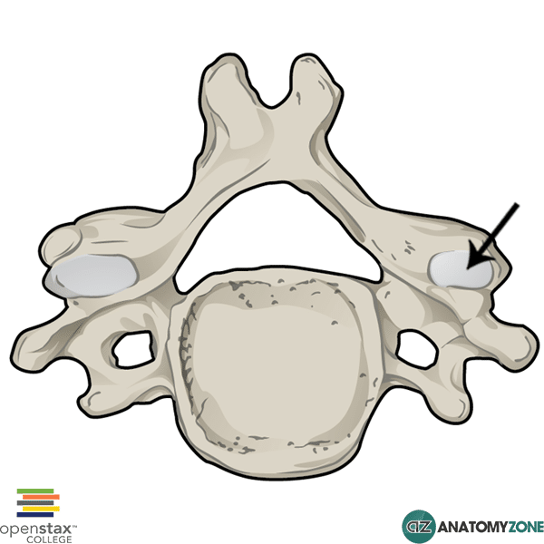
The structure indicated is the superior articular process.

There are 33 vertebrae which make up the spinal column:

  • 7 cervical
  • 12 thoracic
  • 5 lumbar
  • 5 fused sacral vertebrae
  • 3-4 fused coccygeal vertebrae

The typical vertebra consists of the following features:

  • Vertebral body
  • Vertebral arch (neural arch) consisting of lamina and pedicle
  • Spinous process
  • Transverse process
  • Vertebral foramen (collectively form vertebral canal)
  • Superior and inferior articular processes
  • Superior and inferior vertebral notches

The superior articular process articulates with the inferior articular process of the vertebrae above. These articulations are known as zyapophysial joints.

The articular processes arise from the junction between the laminae and pedicles of the vertebral arch.

Learn more about the anatomy of the vertebrae in this tutorial.

Gluteus Maximus

$
0
0

Gluteus Maximus

Gluteus Maximus

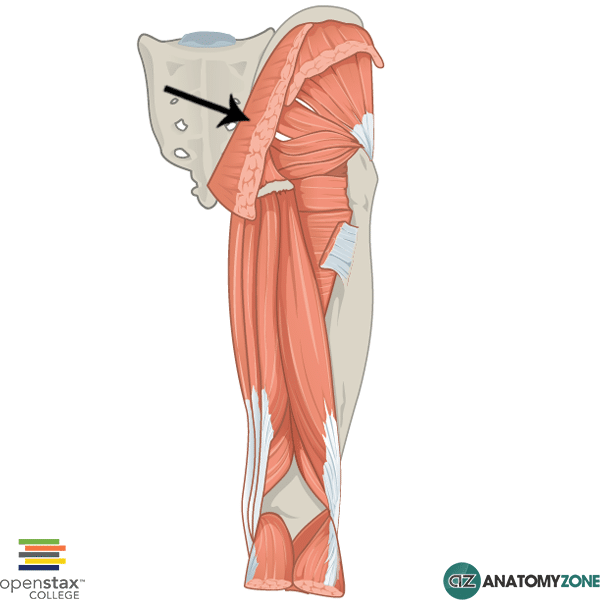
The structure indicated is the gluteus maximus.

The gluteus maximus muscle is one of the muscles of the gluteal region. The muscles in the gluteal region are divided into a superficial and deep group. There are four muscles of the superficial group:

  1. Gluteus maximus
  2. Gluteus medius
  3. Gluteus minimus
  4. Tensor fasciae latae

There are 5 muscles in the deep group:

  1. Gemellus superior
  2. Gemellus inferior
  3. Quadratus femoris
  4. Piriformis
  5. Obturator internus

The gluteus maximus muscle is the largest of the gluteal muscles, and is also the largest muscle in the human body.

Origin: Posterior surface of ilium, posterior superior iliac crest, posterior surface of sacrum and coccyx

Insertion: Primarily to fascia lata at iliotibial tract. Upper posterior surface of femur at gluteal tuberosity.

Action: Hip extension, lateral (external) rotation, abduction (middle and upper fibres)

Innervation: Inferior gluteal nerve

Learn more about the anatomy of the gluteal muscles in this tutorial.

Globus Pallidus

$
0
0

Globus Pallidus

Globus Pallidus

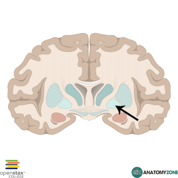
The structure indicated is the globus pallidus.

The globus pallidus is a subcortical structure located within the cerebral hemispheres, and is a major component of the basal ganglia.

The basal ganglia are a group of subcortical nuclei (collection of neuronal cell bodies located within the CNS) located at the base of the forebrain. The term “basal ganglia” is a misnomer, and the name “basal nuclei” would be more appropriate, since ganglia are collections of neuronal cell bodies located within the peripheral nervous system. The basal ganglia form extensive connections with other areas of the brain and are involved mainly in the control of movement and posture.

The basal ganglia consist of the following main components:

  • Caudate nucleus
  • Putamen
  • Globus pallidus
  • Substantia nigra
  • Nucleus accumbens
  • Subthalamic nucleus

These components are grouped together as follows:

  • Caudate nucleus + putamen = striatum
  • Putamen + globus pallidus = lentiform nucleus

The globus pallidus lies medial to the putamen and consists of an internal (medial) and external (lateral) component, separated by the medial medullary lamina. The internal capsule runs medial to the lentiform nucleus. The posterior limb of the internal capsule separates the thalamus from the lentiform nucleus. The anterior limb of the internal capsule separates the putamen from the head of the caudate nucleus.

The globus pallidus is sometimes referred to as the pallidum, or the paleostriatum. The neostriatum is a term used to describe the caudate nucleus and putamen (striatum).

Learn more about the basic anatomy of the brain in this tutorial.

Extensor Digitorum Brevis

$
0
0

 Extensor Digitorum Brevis

 Extensor Digitorum Brevis

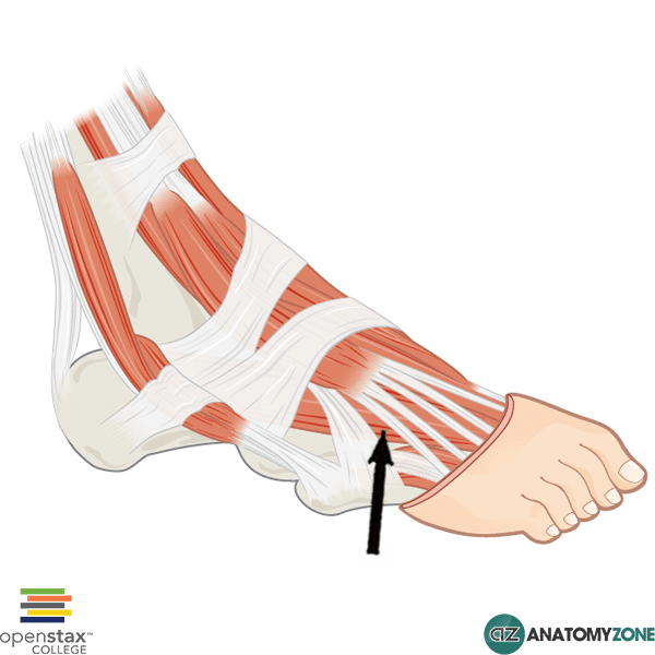
The structure indicated is the extensor digitorum brevis muscle.

This muscle is the only intrinsic muscle of the foot located on the dorsal aspect, and serves to extend digits 2 to 4.

Origin: Superolateral calcaneus

Insertion: Base of proximal phalanx of great toe, lateral portion of tendons of extensor digitorum longus of toes 2 – 4.

Action: Extension of great toe at MTP joint. Flexion of toes 2 – 4.

Innervation: Deep fibular nerve.

Learn more about the anatomy of the foot muscles in this tutorial.

Episode 6 – Control of Tone

$
0
0
Learn all about the physiology of the control of muscle tone in this brand new physiology tutorial.

Flexor Pollicis Brevis

$
0
0

Flexor Pollicis Brevis

Flexor Pollicis Brevis

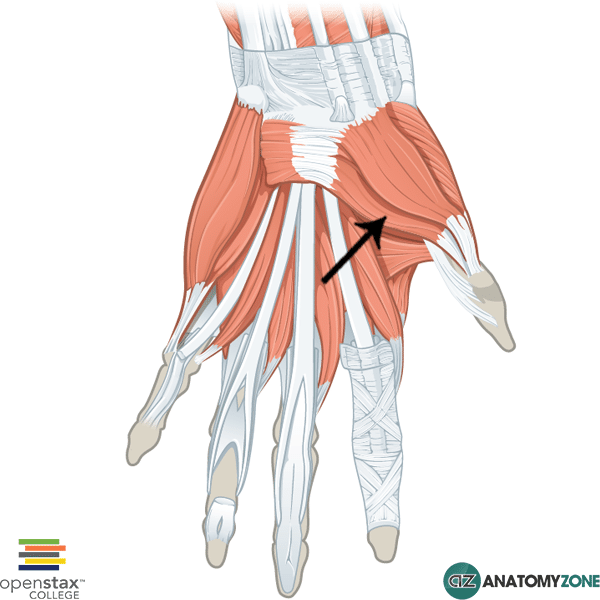
The structure indicated is the flexor pollicis brevis muscle of the hand.

The flexor pollicis brevis muscle is one of the intrinsic muscles of the hand that belongs to the thenar group.

The thenar group of intrinsic hand muscles consist of three muscles:

  • Abductor pollicis brevis
  • Flexor pollicis brevis
  • Opponens pollicis

All the intrinsic muscles of the hand, except the thenar muscles and the lateral two lumbrical muscles are innervated by the deep branch of the ulnar nerve. The thenar muscles and the lateral two lumbrical muscles are innervated by the median nerve.  A useful mnemonic for remembering this is MEATLOAF. “MEAT” refers to the Median nerve, and LOAF refers to the muscles which it innervates: Lateral two lumbricals, Opponens pollicis, Abductor pollicis brevis, Flexor pollicis brevis.

Origin: Flexor retinaculum. Tubercles of scaphoid and trapezium.

Insertion: Lateral base of proximal phalanx of thumb

Innervation: Recurrent branch of median nerve

Action: Flexion of thumb

Learn all about the anatomy of the muscles of the hand in this tutorial.


Cuboid Bone

$
0
0

Cuboid Bone

Cuboid Bone
Cuboid Bone

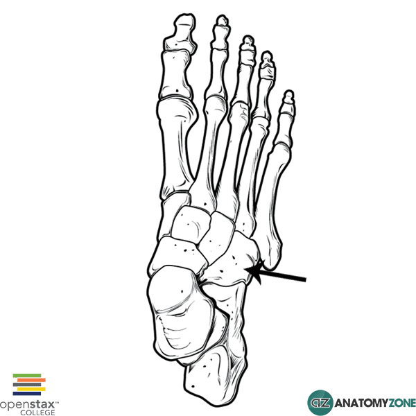
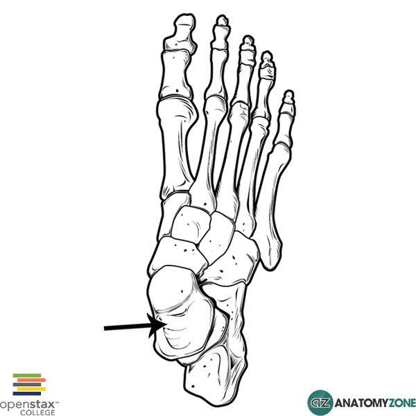
The structure indicated is the cuboid bone of the foot.

The foot contains three groups of bones:

  1. Tarsals
  2. Metatarsals
  3. Phalanges

The cuboid bone is one of seven tarsal bones. There are three groups of tarsal bones:

  • Proximal group
  • Intermediate group
  • Distal group

The cuboid bone belongs to the distal group of tarsal bones. There are four bones in the distal group:

  1. Lateral cuneiform
  2. Intermediate cuneiform
  3. Medial cuneiform
  4. Cuboid

The cuboid bone is the most lateral bone in the distal row of tarsal bones, it is named on the basis of its shape which roughly resembles a cube. The plantar surface of the cuboid bone contains a groove through which the tendon of the peroneus longus travels from the lateral aspect of the foot to insert medially at the base of the first metatarsal and on the medial cuneiform bone. The tibialis posterior muscle inserts onto the plantar surface of the cuboid bone.

Learn more about the bones of the foot in this anatomy tutorial.

First Metacarpal

$
0
0

First Metacarpal

First Metacarpal

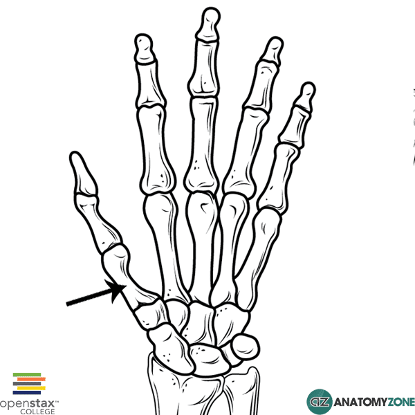
The structure indicated is the first metacarpal of the hand.

There are three groups of bones in the hand:

  1. Carpal bones (8 in total)
  2. Metacarpal bones
  3. Phalanges

The carpal bones are separated into two rows:

  1. Proximal row
  2. Distal row

There are 5 metacarpal bones numbered 1 to 5 and are located between the carpal bones and the phalanges. The first metacarpal bone is associated with the thumb, the 5th metacarpal bone is associated with the little finger. The joints between the carpals and the metacarpals are known as carpometacarpal joints. The joints between the metacarpals and the proximal phalanges are known a metacarpophalangeal (MCP) joints. In addition, the bases of the metacarpal bones articulate with each other.

The first carpometacarpal joint of the thumb is particularly important; the articulation between the first metacarpal and the trapezium bone forms a saddle joint and allows a wide range of movement, including flexion, extension, abduction, adduction, circumduction and opposition.

The metacarpal bones consist of the following parts:

  • Base
  • Shaft
  • Head

Learn more about the anatomy of the bones of the hand in this tutorial.

Adductor Pollicis

$
0
0

Adductor Pollicis

Adductor Pollicis (Oblique Head)
Adductor Pollicis (Transverse Head)

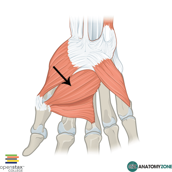
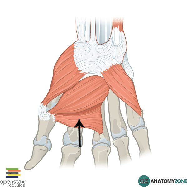
The structure indicated is the adductor pollicis muscle of the hand.

The adductor pollicis muscle belong to the intrinsic group of muscles which act on the hand. The intrinsic muscles include the following muscles/groups:

  • Thenar group (act on thumb)
  • Hypothenar group (act on little finger)
  • Adductor pollicis
  • Lumbricals
  • Interossesus muscles (palmar and dorsal interossei)
  • Palmaris brevis

All the intrinsic muscles of the hand, except the thenar muscles and the lateral two lumbrical muscles are innervated by the deep branch of the ulnar nerve. The thenar muscles and the lateral two lumbrical muscles are innervated by the median nerve.  A useful mnemonic for remembering this is MEATLOAF. “MEAT” refers to the Median nerve, and LOAF refers to the muscles which it innervates: Lateral two lumbricals, Opponens pollicis, Abductor pollicis brevis, Flexor pollicis brevis.

The adductor pollicis muscle consists of two heads:

  • Transverse head
  • Oblique head

As the name suggests, the adductor pollicis serves as a powerful adductor of the thumb. In addition it also assist in opposing the thumb.

Origin: Oblique head – capitate and bases of metacarpals 2 and 3. Transverse head – Metacarpal 3

Insertion: Base of proximal phalanx and extensor expansion of thumb

Action: Adduction of thumb

Innervation: Deep branch of ulnar nerve

Learn more about the anatomy of the hand muscles in this tutorial.

Talus

$
0
0

Talus

Talus
Talus

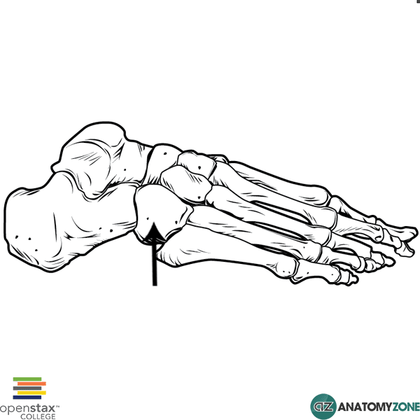
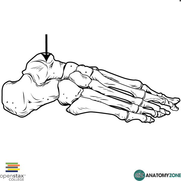
The structure indicated is the talus bone of the foot.

The foot contains three groups of bones:

  1. Tarsals
  2. Metatarsals
  3. Phalanges

The talus is one of seven tarsal bones. There are three groups of tarsal bones:

  1. Proximal group
  2. Intermediate group
  3. Distal group

There are two bones in the proximal group of tarsal bones: the talus and the calcaneus. The calcaneus is the largest tarsal bone and forms the heel through its posterior projection. The talus articulates with the medial malleolus of the tibia and the lateral malleolus of the fibula to form the ankle joint. In addition, it articulates with two other tarsal bones: the calcaneus inferiorly, and the navicular anteriorly.

The talus consists of the following main components:

  • Head: contains facets for articulation with navicular and calcaneus
  • Neck
  • Body: elevated trochlear surface articulates with inferior end of tibia

Learn more about the bones of the foot in this anatomy tutorial.

First Metatarsal

$
0
0

First Metatarsal

First Metatarsal

The structure indicated is the first metatarsal bone of the foot.

The foot contains three groups of bones:

  1. Tarsals
  2. Metatarsals
  3. Phalanges

There are 7 tarsal bones, and 5 metatarsals. The metatarsals are numbered from 1 to 5 from medial to lateral, with the first metatarsal belonging to that of the great toe (hallux) and the 5th metatarsal belonging to that of the little toe.

The metatarsals consist of the following parts:

  • Base
  • Shaft
  • Head

Learn more about the anatomy of the bones of the foot in this anatomy tutorial.

Viewing all 279 articles
Browse latest View live