Quantcast
Channel: AnatomyZone
Viewing all 279 articles
Browse latest View live

Right Coronary Artery

$
0
0

Right Coronary Artery

Right coronary artery

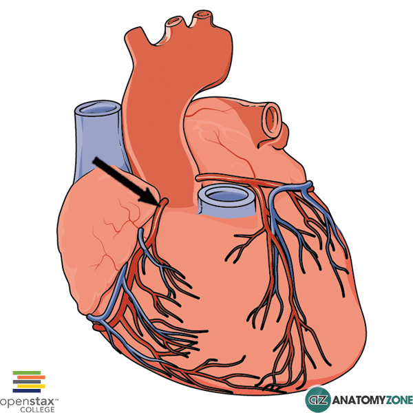
The structure indicated is the right coronary artery of the heart.

The coronary arteries arise from the aortic sinuses of the ascending aorta. The right coronary artery (RCA) arises from the right aortic sinus and the left coronary from the left aortic sinus. The right coronary artery then runs in the coronary sulcus, which separates the atria from the ventricles.

The right coronary artery gives off the following branches:

The coronary artery from which the posterior descending artery arises determines the coronary dominance. The posterior descending artery may arise from the circumflex artery of the left coronary artery. Right dominant is more common than left dominant.

Learn more about the blood supply to the heart in this anatomy tutorial.


Intertubercular sulcus

$
0
0

Intertubercular Sulcus

Intertubercular sulcus

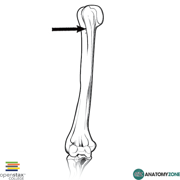
The structure indicated is the intertubercular sulcus of the humerus.

The intertubercular sulcus, also known as the intertubercular groove, or bicipital groove, is a groove separating the greater and lesser tubercles of the humerus.

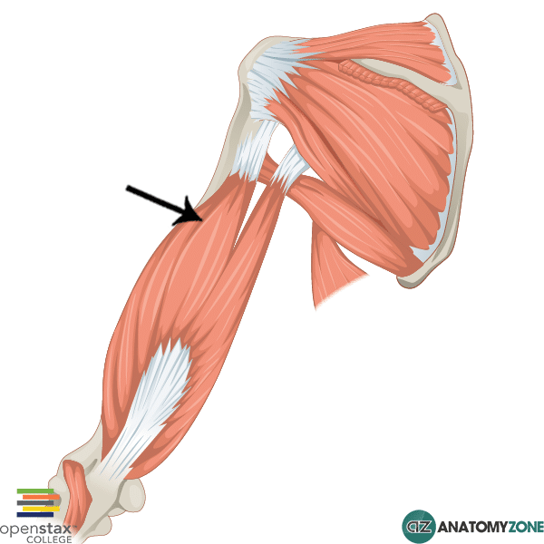
The tendon of the long head of the biceps muscle runs in this groove and attaches on the supraglenoid tubercle of the scapula. The short head of the biceps muscle on the other hand has its origin on the coracoid process of the scapula.

The intertubercular sulcus is the site of three important muscle attachments:

  1. Pectoralis major
  2. Latissmus dorsi
  3. Teres major

The pectoralis major attaches to the lateral lip of the intertubercular sulcus. The latissimus dorsi attaches to the floor of the intertubercular sulcus. The teres major attaches to the medial lip of the intertubercular sulcus. The insertion point of the latissimus dorsi is therefore sandwiched between the insertion points of the teres major and the pectoralis major. This anatomical relationship can be remembered with the mnemonic “the lady between two majors”.

Learn more about the anatomy of the humerus in this tutorial.

Median Sacral Artery

$
0
0

Median Sacral Artery

Median Sacral Artery

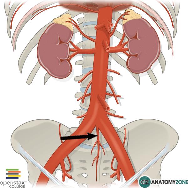
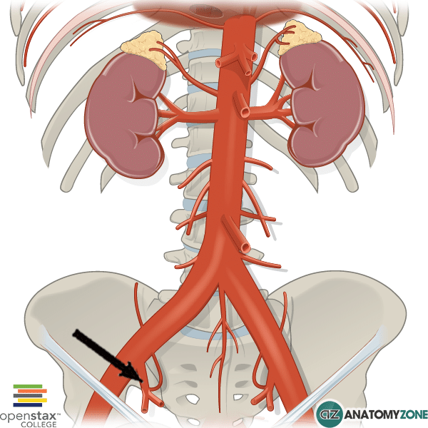
The structure indicated is the median sacral artery.

The median sacral artery arises just above the bifurcation of the aorta into the common iliac arteries. The abdominal aorta gives off anterior, posterior and lateral branches.

The anterior branches include:

  • Celiac trunk
  • Superior mesenteric artery
  • Gonadal arteries
  • Inferior mesenteric arteries

Lateral branches:

  • Middle suprarenal arteries
  • Renal arteries

Posterior branches:

  • Inferior phrenic arteries
  • Lumbar arteries
  • Median sacral arteries

The median sacral artery supplies the coccyx, the lumbar vertebrae and the sacrum.

Lower Limb Veins

$
0
0

Hi this is Peter from AnatomyZone and in this tutorial I’ll be providing you with an overview of the venous drainage of the lower limb.

Read More

The venous drainage of the lower limb consists of a superficial and a deep system. The superficial system is located in the subcutaneous tissue, whereas the deep system is located in the deep fascia of the lower limb. The veins of the deep system accompany the vessels of the arterial system, and they follow a similar naming structure. So if you have a good grasp of the arterial system you’ll be familiar with the names of the deep system of the lower limb. Ultimately, the veins of the superficial system drain to the veins of the deep venous system.


We’ll begin this tutorial by looking at the deep system of veins, and we’ll start distally. Beginning in the foot what we’re looking at here is an anterior view of the foot, and we can see both the venous system and arterial system included on this model. In light blue you can see the vein which accompanies the arcuate artery, and this drains into the vein which accompanies the dorsalis pedis artery, which you can see in green. These deep veins of the foot then drain into the anterior tibial veins, which you can see in the purple colour.


If I rotate the model so we can look at the plantar surface of the foot, you can see that there’s these veins accompanying the deep plantar arterial arch, so this is the deep plantar venous arch, and just like the arterial system, with the medial and lateral plantar arteries, we’ve also got medial and lateral plantar veins. In green we’ve got the lateral plantar vein, and in the light blue colour we’ve got the plantar plantar veins, accompanying the medial plantar artery. These plantar veins then drain into the posterior tibial vein, which you can see runs just behind the medial malleolus and it accompanies the posterior tibial artery.


I’ve just brought the model up, so we’re now looking at a posterior view of the leg, just inferior to the knee joint, and you can see the posterior tibial vein in light blue colour draining into the popliteal vein. And if I rotate the model anteriorly, you can see the anterior tibial veins, which I highlighted in purple colour, draining into the popliteal vein. So it passes from this anterior compartment, to the posterior compartment, and drains into the popliteal vein.


Also on this model, you can see this model highlighted in the green colour. This vein accompanies the fibular artery and is known as the fibular vein, and this vein drains the lateral compartment of the leg.


Now if we follow the course of the popliteal vein, you can see that it follows the course of the popliteal artery, and it passes through the adductor hiatus of the adductor magnus muscle. And it passes from the posterior compartment into the anterior compartment of the thigh. After passing through the hiatus into the anterior compartment, it is then known as the femoral vein. If we follow the femoral vein up proximally, we can see that it receives this tributary, which is the deep vein of the thigh, or the profunda femoris vein. And this vein accompanies the profunda femoris artery. You can also see some other branches which drain into the profunda femoris, and these veins accompany the lateral and medial femoral circumflex arteries.


Just like the perforating arteries of the profunda femoris artery, there are also perforating veins which drain into the deep vein of the thigh. If we follow the femoral vein up even further proximally, we can see that it passes underneath the inguinal ligament. After it passes underneath this ligament, it becomes known as the external iliac vein. The external iliac vein then joins the internal iliac vein to become the common iliac vein. The left and right common iliac veins then unite to form the inferior vena cava.


So you may have heard of a DVT, which stands for deep vein thrombosis. So this is the formation of a clot which forms in the deep system of veins which I’ve just described. And if this clot is dislodged, it can pass into the right side of the heart, through the pathways that I’ve just shown you. So from the deep veins of the leg, into the deep veins of the thigh, and then through the iliac system, into the inferior vena cava and then into the right side of the heart. From the right side of the heart, it can then pass into the pulmonary circulation resulting in a pulmonary infarct. This is known as a pulmonary embolus.


Now moving onto the superficial venous system, this essentially consist of two veins: you’ve got the small saphenous vein and the great saphenous vein. The great saphenous vein you can see here highlighted in light blue, and it runs along the entire length of the leg and the thigh. The small saphenous vein on the other hand, runs posteriorly up the leg and its highlighted here in the green colour. I’ve just moved the model distally and we’re taking a look at the dorsal aspect of the foot. Here we have the dorsal venous arch, which drains the dorsal aspect of the foot and on the plantar aspect of the foot is a venous network, which drains the plantar structures. Coming back to the dorsal view, if we just rotate around to a lateral view, we can see the small saphenous vein arising from the lateral aspect of the dorsal venous arch and passing behind the lateral malleolus to ascend the leg. The small saphenous vein then ascends posteriorly up the leg to the level of the knee and it then drains into the popliteal vein behind the knee joint. I’ve just removed the structures so you can see the small saphenous vein draining into the popliteal vein, and this is a point where the superficial system meets the deep venous system.


Coming back distally to the foot and taking a look at the medial aspect of the dorsal venous arch, we can see the great saphenous vein given off. We can see that the great saphenous vein passes in front of the medial malleolus and then it runs along the medial aspect of the leg, along the entire length of the lower limb. It then drains into the deep venous system by draining into the femoral vein, which we took a look at before.


In addition to these two major veins, the small and great saphenous vein, you also have perforating veins. These are small little veins which pass directly from the superficial venous system to the deep venous system. So the deep venous system is actually at a higher pressure to the superficial venous system. In order to prevent the backflow of blood from the deep to the superficial system, there are valves at these junctions between the saphenous vein and the deep system. So where the great saphenous vein drains into the femoral vein, you have a valve which prevents that higher pressure blood flowing back into the superficial, and likewise, at the popliteal vein where the small saphenous vein drains into the popliteal, you have a valve to prevent the backflow of blood into the superficial small saphenous vein.


So if these valves become incompetent, the superficial veins dilate up and take on this tortuous appearance, because of the back flow of blood, this appearance is what is referred to as a varicose vein.


So that’s an overview of the venous system of the lower limb.
Upper Limb Arteries

$
0
0
Learn all about the anatomy of the arteries of the upper limb in this tutorial.

Biceps Femoris

$
0
0

Biceps Femoris

Biceps Femoris

The structure indicated is the biceps femoris muscle of the thigh.

The biceps femoris muscle is one of three muscles in the posterior compartment of the thigh. The other muscles are the semimembranosus, and the semitendinosus.

The muscles in the posterior compartment are often referred to as the “hamstrings” muscles. Collectively, these muscles are responsible for extending the hip joint, and flexing the knee joint.

The biceps femoris lies laterally, and the semitendinosus and semimembranosus lie medially.

Just like the biceps muscle in the arm, the biceps brachii , which is Latin for “two headed muscle (biceps) of the arm (brachii)”, the biceps femoris has two heads – a short head and a long head. The long head originates from the ischial tuberosity together with the semitendinosus muscle, whereas the short head originates from the shaft of the femur on the linear aspera. The two heads converge to a form a tendon which inserts laterally on the fibular head. The prime function of the biceps femoris is in flexion of the knee.

Origin: Long head – ischial tuberosity. Short head – linea aspera

Insertion: Fibula head and lateral tibial condyle

Action: Flexion of knee, lateral rotation of tibia, extension of hip.

Innervation: long head – tibial nerve. Short head – common peroneal nerve.

You can learn more about the muscles of the posterior compartment of the thigh in this video.

Greater Tubercle of Humerus

$
0
0

Greater Tubercle of Humerus

Greater tubercle of humerus

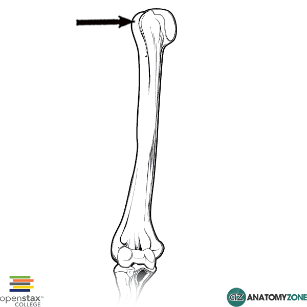
The structure indicated is the greater tubercle of the humerus.

The greater tubercle lies lateral to the head of the humerus as serves as the point of attachment for three of the rotator cuff muscles. There are three flat facets to which these three muscles attach:

  1. Superior facet
  2. Middle facet
  3. Inferior facet

The supraspinatus attaches to the superior facet.

The infraspinatus attaches to the middle facet.

The teres minor attaches to the inferior facet.

Separating the greater tubercle from the lesser tubercle is the intertubercular sulcus, also known as the bicipital groove. The long head of the biceps runs in this groove to attach to the supraglenoid tubercle.

Learn more about the anatomy of the humerus in this anatomy tutorial.

Palmaris Longus

$
0
0

Palmaris Longus

Palmaris Longus

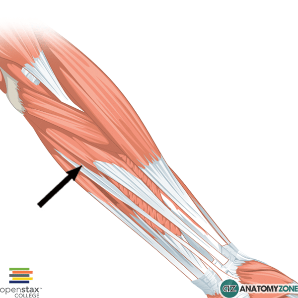
The structure indicated is the palmaris longus muscle of the forearm.

The palmaris longus is one of four muscles in the superficial layer of muscles in the anterior compartment of the forearm. The anterior compartment of the forearm consists of three layers of muscles:

  1. Superficial layer
  2. Intermediate layer
  3. Deep layer

Four muscles are located in the superficial layer (from lateral to medial):

  1. Flexor carpi ulnaris
  2. Palmaris longus
  3. Flexor carpi radialis
  4. Pronator teres

All four muscles of the superficial layer have a common origin on the medial epicondyle of the humerus.

Origin:  medial epicondyle of humerus

Insertion: distal half of flexor retinaculum and palmar aponeurosis

Action: flexion of wrist. Tightens palmar aponeurosis

Innervation: Median nerve

Learn more about the anatomy of the muscles of the forearm in this tutorial.


Obturator Foramen

$
0
0

Obturator Foramen

Obturator Foramen

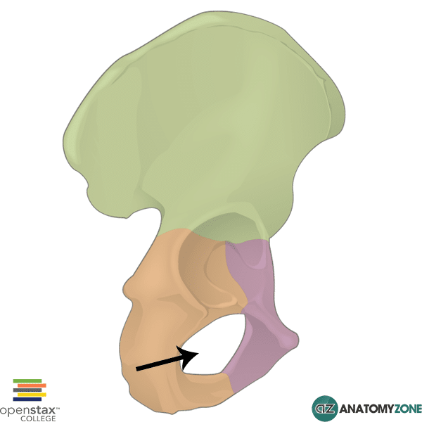
The obturator foramen is indicated in this diagram.

The obturator foramen lies inferior to the acetabulum of the pelvis and is an opening between the ischium and pubic bones. It is covered almost entirely by the obturator membrane. A small gap is left between the superior margin of the obturator membrane and the pelvic bone above, known as the obturator canal, which allows a few vessels and nerves to pass out from the pelvic cavity to communicate with the lower limb. There are three structures which pass through the obturator canal:

  1. Obturator artery
  2. Obturator vein
  3. Obturator nerve

Learn more about the anatomy of the pelvic bones in this tutorial

Temporalis Muscle

$
0
0

Temporalis Muscle

Temporalis Muscle

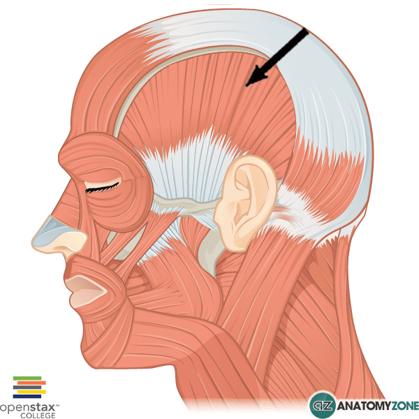
The structure indicated is the temporalis muscle.

This muscle is one of the muscles of mastication. The muscles of mastication include:

  • Masseter
  • Temporalis
  • Medial pterygoid
  • Lateral pterygoid

The temporalis muscle is a large fan-shaped muscle which lies in the temporal fossa above the zygomatic arch.

Origin: temporal line of parietal bone. Temporal surface of sphenoid bone

Insertion: coronoid process of mandible and ramus of mandible

Innervation: mandibular branch of trigeminal nerve (cranial nerve V)

Action: Elevation of the mandible and retraction of the mandible (via posterior horizontal fibres)

To learn more about the muscles of mastication, check out this tutorial!

Patellar ligament

$
0
0

Patellar Ligament

Patellar Ligament

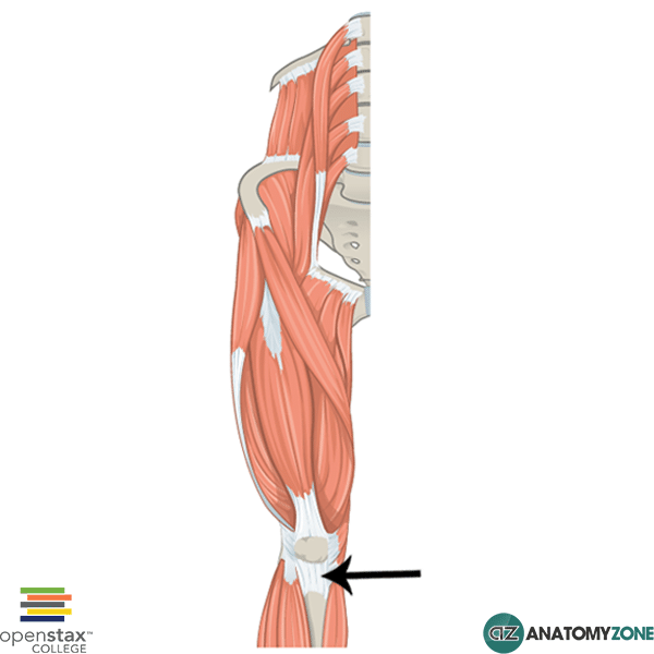
The structure indicated is the patellar ligament.

The quadriceps muscles of the thigh attach to and encapsulate the patella (the kneecap), via the quadriceps tendon. The patella ligament is essentially the continuation of the quadriceps tendon inferior to the patella, attaching it to the tibial tuberosity.

Learn more about the muscles of the thigh in this tutorial.

Tensor Fasciae Latae Muscle

$
0
0

Tensor Fasciae Latae Muscle

Tensor Fasciae Latae Muscle

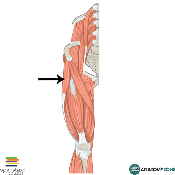
The structure indicated is the tensor fasciae latae muscle.

The tensor fasciae latae muscle is one of the muscles of the gluteal region. The muscles in the gluteal region are divided into a superficial and deep group. There are four muscles of the superficial group:

  1. Gluteus maximus
  2. Gluteus medius
  3. Gluteus minimus
  4. Tensor fasciae latae

There are 5 muscles in the deep group:

  1. Gemellus superior
  2. Gemellus inferior
  3. Quadratus femoris
  4. Piriformis
  5. Obturator internus

The tensor fasciae latae muscle is the most anteriorly situated muscle of the gluteal region and is continuous with the iliotibial tract (iliotibial band), which is the fibrous tract of tissue formed from the lateral thickening of the deep fascia of the thigh. The iliotibial tract attaches to the lateral condyle of the tibia to provide lateral stability to the knee joint. Both the gluteus maximus and the tensor fasciae latae insert onto the iliotibial tract. The deep fascia of the thigh is the “fascia lata” – the literal Latin translation. The name “tensor fasciae latae” therefore comes from the function of this muscle in tensing the fascia lata/deep fascia.

Origin: Anterior superior iliac spine and outer edge of the iliac crest.

Insertion: Iliotibial band

Action: Hip abduction, medial rotation, assists hip flexion, stabilises hip and knee by placing tension in iliotibial band.

Innervation: Superior gluteal nerve

Learn more about the anatomy of the gluteal muscles in this tutorial.

Purkinje Fibres

$
0
0

Purkinje Fibres

Purkinje Fibres

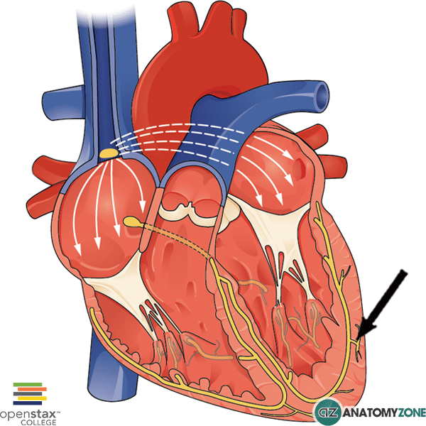
The diagram indicates purkinje fibres.

The conduction system of the heart is responsible for the synchronised contraction of the atria and ventricles and is comprised of the following main structures:

The atrioventricular nodes receives signals from the sinoatrial node. The impulses from the atrioventricular node then pass to the Bundle of His and then follow the left and right bundle branches through the interventricular septum and ultimately to the Purkinje fibres which propagate the signal into the ventricles.

Purkinje fibres are located on the inner aspect of the ventricular walls beneath the endocardium in the subendocardium. These fibres have special properties which allow them to conduct action potentials with great efficiency. They are important in ensuring synchronised ventricular contraction. They extend from the interventricular septum, through the papillary muscles and laterally up the walls of the venricles.

Internal Iliac Artery

$
0
0

Internal Iliac Artery

Internal Iliac Artery

The structure indicated is the internal iliac artery.

The abdominal aorta bifurcates into the left and right common iliac arteries at approximately the level of the fourth lumbar vertebra. The common iliac arteries then divide into the external and internal iliac arteries.

The internal iliac artery has an anterior trunk and posterior trunk. The branches of these trunks are anatomically variable and have several different configurations from person to person. The posterior trunk gives rise to three branches:

  1. Iliolumbar artery
  2. Lateral sacral arteries
  3. Superior gluteal artery

The branches of the anterior trunk are much more variable, but tend to give rise to the following arteries:

  1. Umbilical artery – gives rise to superior vesical artery
  2. Obturator artery
  3. Inferior vesical artery (vaginal artery in women)
  4. Middle rectal artery
  5. Internal pudendal artery
  6. Inferior gluteal artery
  7. Uterine artery (in women)

The internal iliac artery is the main artery of the pelvis and supplies the pelvic walls and the viscera that are contained within the pelvis, as well as the musculature of the gluteal region and muscles located medially in the thigh.

Learn all about the anatomy of the internal iliac artery in this anatomy tutorial.

Acetabulum

$
0
0

Acetabulum

Acetabulum

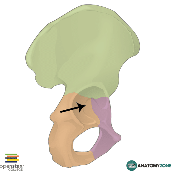
The structure indicated is the acetabulum of the pelvis.

The acetabulum is formed from the point joining the three bones of the pelvis (ilium, ischium and pubis), and is the site of articulation with the head of the femur. The femoroacetabular articulation is known as the hip joint.

The acetabulum consists of two parts:

  • Articular part
  • Non-articular part

The non-articular part consists of the acetabular fossa and the acetabular notch inferiorly. The ligamentum teres attaches at one end to the acetabular notch, and at the other end to the fovea capitis of the femur. The articular part forms a rim surrounding the acetabular fossa, serving as a point of attachment for the acetabular labrum. This labrum is a ring of cartilage which functions to deepen the cavity of the acetabulum, stabilising the hip joint and making it structurally more difficult for the head of the femur to dislodge from its acetabular articulation.

Learn more about the hip joint, pelvis and femur in these anatomy tutorials.


Lateral Head of Triceps Brachii

$
0
0

Lateral Head of Triceps Brachii

Lateral Head of Triceps Brachii

The structure highlighted is the lateral head of the triceps brachii.

The triceps brachii is the sole muscle in the posterior compartment of the arm. As the name suggests, the triceps brachii has three heads:

  1. Long head
  2. Medial head
  3. Lateral head

These three heads converge to insert on the olecranon of the ulna. The lateral head and long head of the triceps brachii lie superficially, the medial head lies deep to these two heads. The triceps muscle serves to extend the forearm at the elbow joint and is an antagonist of the biceps and brachialis muscles of the anterior compartment of the arm.

Origin:

  • Long head – infraglenoid tubercle of scapula
  • Medial head – posteriorly on the shaft of the humerus below the radial groove
  • Lateral head – posteriorly on the humerus, above the radial groove

Insertion: Olecranon of the ulna

Action: Extension of the forearm at the elbow joint

Innervation: Radial nerve

Learn more about the anatomy of the arm muscles in this anatomy tutorial.

Adductor Longus

$
0
0

Adductor Longus

Adductor Longus

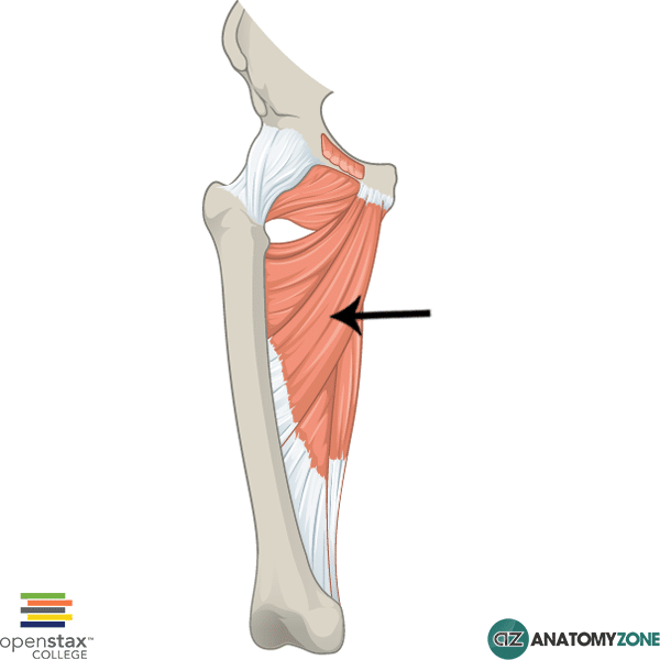
The muscle indicated is the adductor longus muscle of the thigh.

The adductor longus is one of six muscles in the medial compartment of the thigh. The thigh consists of three muscular compartments:

  1. Anterior
  2. Medial
  3. Posterior

The medial compartments consists of the following muscles:

  • Gracilis
  • Pectineus
  • Adductor longus
  • Adductor brevis
  • Adductor magnus
  • Obturator externus

All the muscles of the medial compartment are innervated by the obturator nerve except for the pectineus (femoral nerve) and the hamstring part of the adductor magnus (tibial division of sciatic). The muscles of the medial compartment generally serve to adduct the thigh at the hip joint.

The adductor longus can additionally medially rotate the femur. There are three “adductor” named muscles: adductor brevis, adductor longus and adductor magnus. The adductor brevis and longus lie superficial to the adductor magnus. The adductor brevis is located superiorly and the adductor longus is located inferiorly.

Origin: anterior body of pubis

Insertion: middle third of linea aspera

Action: adduction, medial rotation and flexion of thigh at hip joint.

Innervation: obturator nerve

Learn more about the anatomy of the thigh muscles in this anatomy tutorial.

Left Gonadal Vein

$
0
0

Left Gonadal Vein

Left Gonadal Vein

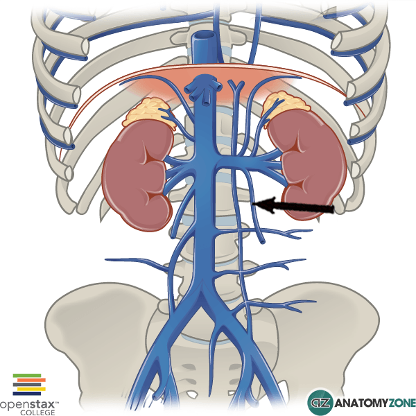
The structure indicated is the left gonadal vein.

The gonadal veins are the veins which drain the testes in men (testicular vein) and the ovaries (ovarian vein) in women.  The left gonadal vein drains into the left renal vein, which then drains into the inferior vena cava. The right gonadal vein however, drains directly into the inferior vena cava.

The inferior vena cava is responsible for carrying deoxygenated blood into the right atrium of the heart from the lower part of the body. The inferior vena cava is formed at the point of union of the right and left common iliac veins at the L5 vertebral level.

Several veins drain into the inferior vena cava:

  • Lumbar veins
  • Right gonadal vein (the left gonadal vein drains into the left renal vein)
  • Renal veins
  • Right suprarenal vein
  • Inferior phrenic vein
  • Hepatic veins

Learn more about the veins of the body in this tutorial.

Adductor Tubercle

$
0
0

Adductor Tubercle

Adductor Tubercle

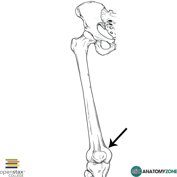
The structure indicated is the adductor tubercle of the distal femur.

The adductor tubercle is found distally on the femur and is formed from the termination of the medial supracondylar line. The adductor tubercle serves as the point of attachment for the adductor magnus muscle.

Learn more about the anatomy of the femur in this tutorial.

Proximal Phalanx

$
0
0

Proximal Phalanx

Proximal Phalanx

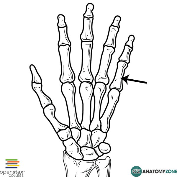
The structure indicated is the proximal phalanx of the 5th finger.

The hand consists of three groups of bones:

  1. Carpals
  2. Metacarpals
  3. Phalanges

The phalanges (phalanx = singular), form the fingers/digits of the hands. The thumb consists of two phalanges: proximal phalanx and distal phalanx. The rest of the fingers consist of:

  • Proximal phalanx
  • Middle phalanx
  • Distal phalanx

The phalanges themselves have the following features:

  • Base (proximally)
  • Head (distally)
  • Shaft/body (in between the base and the head)

Learn more about the anatomy of the bones of the hand in this tutorial.

Viewing all 279 articles
Browse latest View live