Quantcast
Channel: AnatomyZone
Viewing all 279 articles
Browse latest View live

Lesser Trochanter

$
0
0

Lesser Trochanter

Lesser Trochanter
Lesser Trochanter

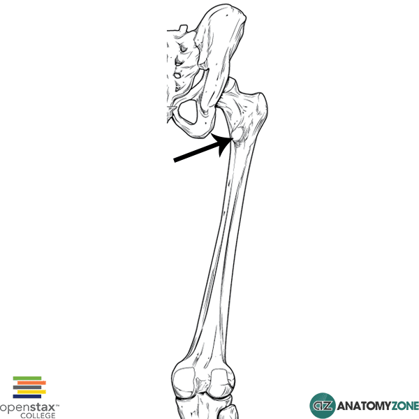
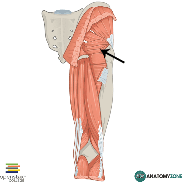
The structure indicated is the lesser trochanter of the femur.

The lesser trochanter is a small protuberance of bone that projects from the posterior aspect of the femur, inferomedially at the base of the femoral neck. Two muscles insert onto the lesser trochanter:

Learn more about the anatomy of the femur in this anatomy tutorial.


Upper Limb Arteries – Hand and Wrist

$
0
0
Learn all about the anatomy of the arteries of the upper limb in this tutorial.

Pulmonary Trunk

$
0
0

Pulmonary Trunk

Pulmonary Trunk

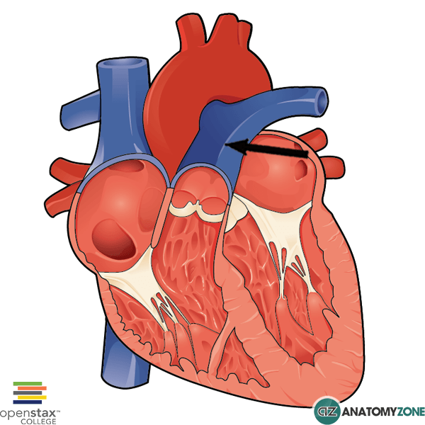
The structure indicated is the pulmonary trunk.

The pulmonary trunk is one of the great vessels – a term which refers to the major arteries and veins which bring blood to and from the heart. These include:

  • Superior and inferior vena cavae
  • Pulmonary arteries
  • Pulmonary veins
  • Aorta

The pulmonary trunk carries deoxygenated blood from the right ventricle of the heart to the lungs for oxygenation, via the right and left pulmonary arteries. This is the only artery in the adult human body that carries deoxygenated blood. In fetal life, the umbilical arteries also carry deoxygenated blood – this artery extends into the umbilical cord and to the placenta for oxygenation.

The pulmonary trunk bifurcates to form the left and the right pulmonary arteries. The right pulmonary artery passes underneath the arch of the aorta towards the right lung, and the left pulmonary artery extends to the left lung.

Learn more about the great vessels in this tutorial

Adductor Brevis

$
0
0

Adductor Brevis

Adductor Brevis

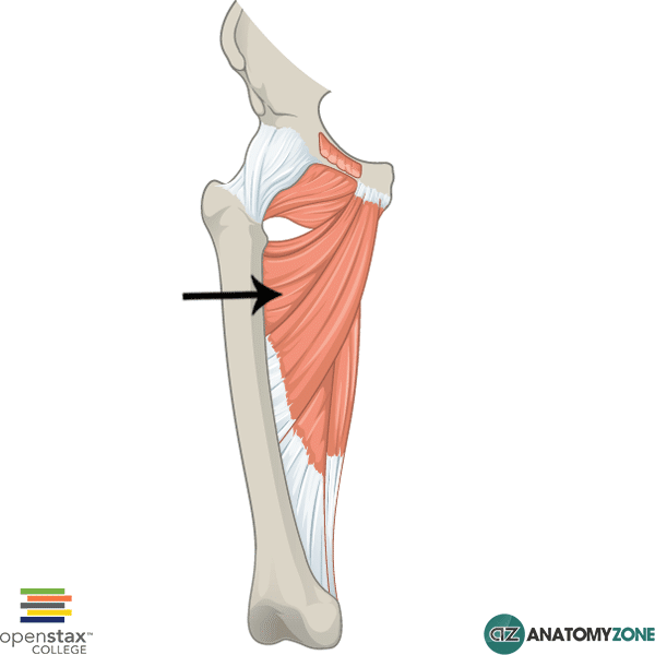
The structure indicated is the adductor brevis muscle.

The muscle indicated is the adductor brevis muscle of the thigh.

The adductor brevis is one of six muscles in the medial compartment of the thigh. The thigh consists of three muscular compartments:

  1. Anterior
  2. Medial
  3. Posterior

The medial compartments consists of the following muscles:

All the muscles of the medial compartment are innervated by the obturator nerve except for the pectineus (femoral nerve) and the hamstring part of the adductor magnus (tibial division of sciatic). The muscles of the medial compartment generally serve to adduct the thigh at the hip joint.

There are three “adductor” named muscles: adductor brevis, adductor longus and adductor magnus. The adductor brevis and longus lie superficial to the adductor magnus. The adductor brevis is located superiorly and the adductor longus is located inferiorly. The adductor brevis lies deep to the adductor longus and pectineus muscles.

Origin: anteroinferior pubic ramus

Insertion: pectineal line, superomedial part of linea aspera

Action: adducts hip. Also lateral rotation of hip and flexion of hip.

Innervation: obturator nerve

Learn more about the anatomy of the thigh muscles in this anatomy tutorial.

Obturator Externus

$
0
0

Obturator Externus

Obturator Externus

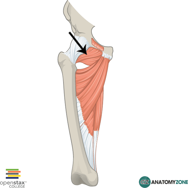
The muscle indicated is the obturator externus muscle.

The obturator externus is one of six muscles in the medial compartment of the thigh. The thigh consists of three muscular compartments:

  1. Anterior
  2. Medial
  3. Posterior

The medial compartments consists of the following muscles:

All the muscles of the medial compartment are innervated by the obturator nerve except for the pectineus (femoral nerve) and the hamstring part of the adductor magnus (tibial division of sciatic). The muscles of the medial compartment generally serve to adduct the thigh at the hip joint.

There are two obturator muscles: obturator externus and obturator internus. The obturator muscles are attached to the obturator membrane which covers the obturator foramen of the pelvic bone. The obturator externus as the name suggests is located on the external surface of the obturator membrane and belongs to the medial compartment of thigh muscles. The obturator internus is located internally on the pelvis and belongs to the deep group of gluteal muscles.

Origin: obturator membrane, anterior obturator foramen

Insertion: posteriomedial surface of greater trochanter of femur in lateral wall of trochanteric fossa

Action: obturator nerve

Innervation: lateral rotation of hip. Assists hip adduction.

Learn more about the anatomy of the thigh muscles in this anatomy tutorial.

Calcaneal (Achilles) Tendon

$
0
0

Calcaneal (Achilles) Tendon

Calcaneal (Achilles) Tendon

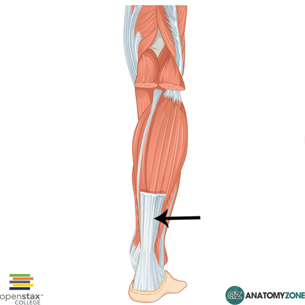
The structure indicated is the calcaneal tendon (Achilles’ tendon).

The calcaneal tendon is formed from the convergence of the three muscles of the superficial compartment of the posterior leg to form a tendon that inserts onto the calcaneus. The posterior compartment of the leg consists of a superficial group of muscles and a deep group of muscles.

The superficial group consists of the following leg muscles:

  • Gastrocnemius
  • Plantaris
  • Soleus

The deep group consists of the following leg muscles:

  • Popliteus
  • Flexor hallucis longus
  • Flexor digitorum longus
  • Tibialis posterior

The calcaneal tendon is the thickest tendon in the human body. Contraction of the muscles attaching via this tendon results in plantarflexion of the foot at the ankle joint. The calcaneal tendon gets the name “Achilles’ tendon” from Greek mythology whereby the Greek hero Achilles was killed by Paris of Troy in the Trojan war by a poisoned arrow to the heel.

Learn more about the anatomy of the leg muscles in this tutorial.

Greater Sciatic Notch

$
0
0

Greater Sciatic Notch

Greater Sciatic Notch

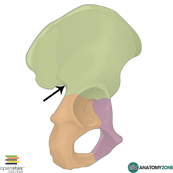
The structure indicated is the greater sciatic notch of the pelvis.

The greater sciatic notch les between the ischial spine (inferiorly) and the posterior inferior iliac spine (superiorly). The sacrospinous ligament which attaches from the ischial spine to the lateral aspect of the sacrum and coccyx converts the greater sciatic notch into the greater sciatic foramen.

The lesser sciatic notch lies below the ischial spine, and is converted into the lesser sciatic foramen by the sacrospinous and sacrotuberous ligaments.

The piriformis muscle passes through the greater sciatic foramen and divides it into two spaces:

  • Greater sciatic foramen above the piriformis (suprapiriform foramen)
  • Greater sciatic foramen below the piriformis (infrapiriform foramen)

The following structures pass in the foramen above the piriformis:

  • Superior gluteal vessels
  • Superior gluteal nerve

The following structures pass in the foramen below the piriformis:

  • Inferior gluteal vessels
  • Internal pudendal vessels
  • Inferior gluteal nerve
  • Pudendal nerve
  • Sciatic nerve
  • Posterior femoral cutaneous nerve
  • Nerve to obturator internus
  • Nerve to quadratus femoris

Learn about the bones of the pelvis in this anatomy tutorial.

Radial Fossa

$
0
0

Radial Fossa

Radial Fossa

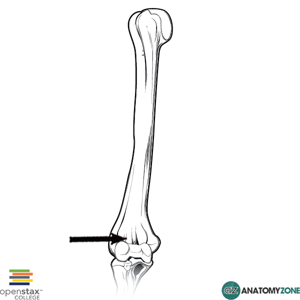
The structure indicated is the radial fossa of the distal humerus.

The distal end of the humerus consists of several features:

  • Condyle, consisting of the capitulum and trochlea
  • Medial and lateral epicondyles
  • Medial and lateral supracondylar ridges
  • Radial fossa, coronoid fossa, olecranon fossa

A large central condyle which has two articular components – the capitulum which articulates with the radius, and the trochlea which articulates with the ulna. Either side of the humeral condyle, are two epicondyles, the medial and lateral epicondyles superior to which are the medial and lateral supracondylar ridges.

There are three fossae found distally on the humerus:

  1. Radial fossa
  2. Coronoid fossa
  3. Olecranon fossa

The radial fossa is located just above the capitulum, the coronoid fossa is located just above the trochlea. The olecranon fossa is located posteriorly on the humerus above the trochlea. When the forearm is flexed, the radial fossa accommodates the anterior aspect of the radial head, facilitating movement. Likewise, the coronoid fossae accommodates the coronoid process of the ulna during flexion of the forearm. The olecranon fossa, as the name suggests accommodates the apex of the olecranon when the elbow joint is in extension.

Learn more about the anatomy of the humerus in this anatomy tutorial.


Superior Angle of Scapula

$
0
0

Superior Angle of Scapula

Superior Angle of Scapula

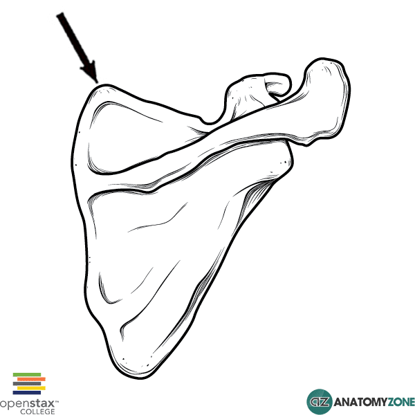
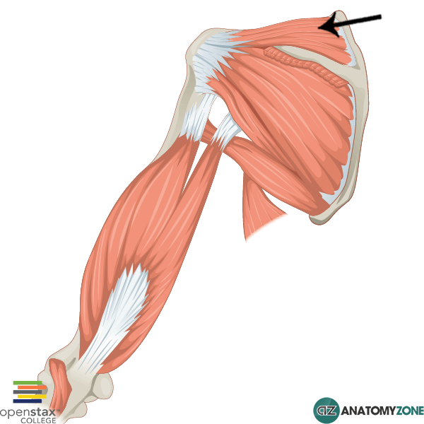
The superior angle of the scapula is indicated in the diagram.

The scapula is a bone that connects the humerus with the clavicle. The scapula consists of two surfaces:

  1. Costal
  2. Posterior

The scapula has three borders:

  1. Superior
  2. Medial
  3. Lateral

The scapula has three angles:

  1. Superior
  2. Inferior
  3. Lateral

The superior angle of the scapula is covered by the trapezius muscle. It is formed at the point where the medial and lateral borders meet and provides attachment for the levator scapulae muscle and omohyoid muscle.

Learn more about the anatomy of the scapula in this tutorial

Flexor Hallucis Brevis

$
0
0

Flexor Hallucis Brevis

Flexor Hallucis Brevis

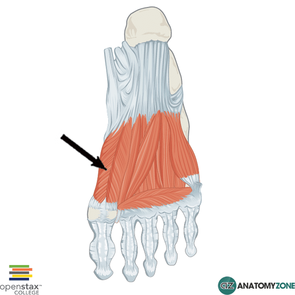
The structure indicated is the flexor hallucis brevis muscle of the foot.

The intrinsic muscles on the plantar aspect of the foot are divided into four layers. The flexor hallucis brevis muscle is located in the third layer of intrinsic foot muscles. There are three muscles located in this layer:

  1. Flexor hallucis brevis
  2. Adductor hallucis
  3. Flexor digiti minimi brevis

The flexor hallucis brevis muscle splits into two parts which insert onto the medial and lateral aspects of the base of the first phalanx of the great toe.

Origin: Plantar surface of cuboid and lateral cuneiform bone

Insertion: Lateral and medial sides of base of proximal phalanx of the great toe

Action: Flexes great toe at metatarsophalangeal joint

Innervation: medial plantar nerve

Learn more about the anatomy of the foot muscles in this tutorial.

Lateral Pterygoid

$
0
0

Lateral Pterygoid

Lateral Pterygoid Muscle

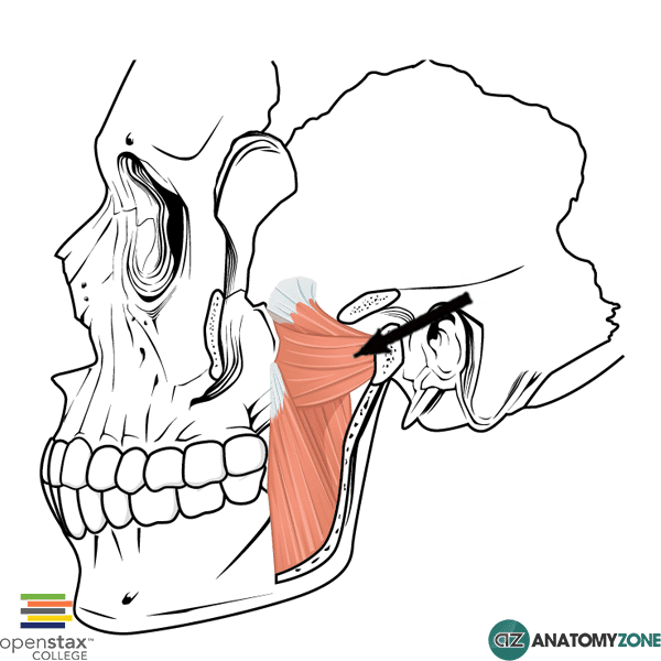
The structure indicated is the lateral pterygoid muscle.

The lateral pterygoid muscle is one of four muscles of mastication:

  1. Masseter
  2. Temporalis
  3. Medial pterygoid
  4. Lateral pterygoid

The muscles of mastication act on the temporomandibular joint (TMJ), which is the joint of the jaw formed between the mandible and temporal bone. The TMJ is a synovial joint, which differs in structure to other synovial joints in that it contains a fibrocartilagenous articular disc that divides the joint into two parts. The two joint compartments result in different movements at the TMJ. The lower joint compartment allows rotational movement, whilst the upper joint compartment allows translational movement. There are four movements at the TMJ:

  1. Protrusion
  2. Retraction
  3. Elevation
  4. Depression

The lateral pterygoid muscle is located in the infratemporal fossa and consists of two heads:

  • Upper head
  • Lower head

Due to the primarily horizontal orientation of the fibres of the lateral pterygoid, its primary function is protrusion of the mandible.

Origin: Superior head: infratemporal crest of greater winger of sphenoid. Inferior head: lateral plate of the pterygoid process

Insertion: Conjoined tendon to neck of mandible

Action: bilateral contraction: protrusion of mandible. Unilateral contraction: side-side jaw movements

Innervation: mandibular branch of trigeminal nerve (cranial nerve V)

Learn more about the anatomy of the muscles of mastication in this anatomy tutorial.

Latissimus Dorsi

$
0
0

Latissimus Dorsi

Latissimus Dorsi

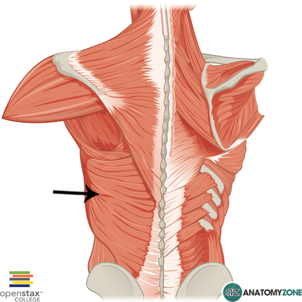
The structure indicated is the latissimus dorsi muscle.

The latissimus dorsi muscle is one of the superficial muscles of the back. There are three groups of back mucsles:

  1. Superficial
  2. Intermediate
  3. Deep

The superficial group of back muscles consists of 5 muscles:

  1. Trapezius
  2. Latissimus dorsi
  3. Levator scapulae
  4. Rhomboid major
  5. Rhomboid minor

“Latissimus dorsi” translates literally from the Latin has the “broadest muscle of the back”, which reflects its large, flat structure. The latissimus dorsi muscle is involved in several movements of the upper limbs as well as movement at the lumbar spine and it also plays a role as an accessory muscle of inspiration.

Origin: Spinous processes of T7-L5, sacrum, iliac crest, ribs 10-12

Insertion: Bicipital groove of humerus

Action: extension, adduction and medial rotation of humerus

Innervation: thoracodorsal nerve

Learn more about the anatomy of the back muscles in this tutorial.

Flexor Carpi Radialis

$
0
0

Flexor Carpi Radialis

Flexor Carpi Radialis

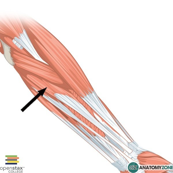
The structure indicated is the flexor carpi radialis muscle.

The flexor carpi radialis is one of four muscles in the superficial layer of muscles in the anterior compartment of the forearm. The anterior compartment of the forearm consists of three layers of muscles:

  1. Superficial layer
  2. Intermediate layer
  3. Deep layer

Four muscles are located in the superficial layer (from lateral to medial):

  1. Flexor carpi ulnaris
  2. Palmaris longus
  3. Flexor carpi radialis
  4. Pronator teres

All four muscles of the superficial layer have a common origin on the medial epicondyle of the humerus.

Origin: Medial epicondyle of humerus

Insertion: Bases of 2nd and 3rd metacarpals

Action: flexion and abduction of wrist

Innervation: median nerve

Learn more about the anatomy of the forearm muscles in this tutorial.

Superior Extensor Retinaculum

$
0
0

Superior Extensor Retinaculum

Superior Extensor Retinaculum

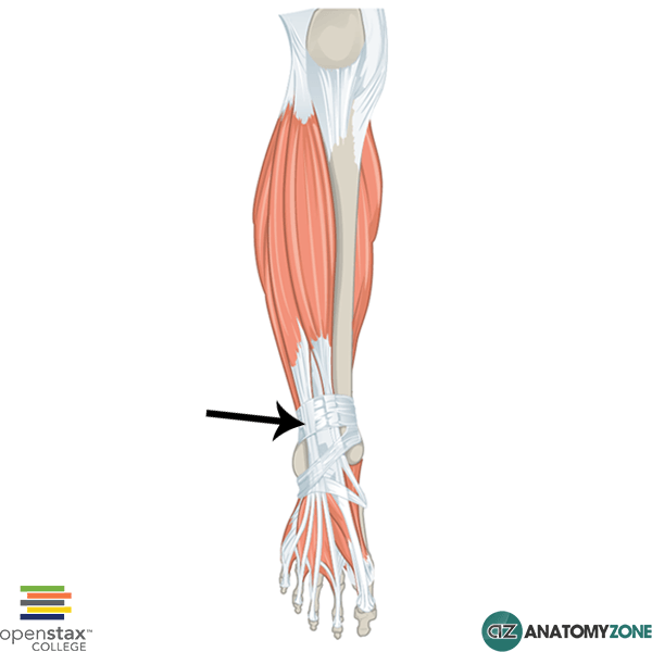
The structure indicated is the superior extensor retinaculum of the leg.

Retinacula (retinaculum singular) are bands of connective tissue which surround tendons and hold them in place. They function to stabilise tendons as the muscles to which they attach contract to cause movement. There are retinacula in the wrist, ankle and knee.

In the ankle, there is the flexor retinaculum, the peroneal retinacula and on the extensor surface, there is the superior and inferior extensor retinacula. The superior retinaculum is situated distally in the leg just above the ankle joint, attaching to the anterior aspects of the fibula and tibia.

The inferior retinaculum is Y-shaped. The base of the “Y” attaches laterally on the calcaneus. The upper arm of the “Y” attaches to the medial malleolus, whereas the lower arm of the “Y” attaches medially on the plantar aponeurosis.

The following tendons pass underneath the superior extensor retinaculum:

  • Extensor digitorum longus tendon
  • Extensor hallucis longus tendon
  • Peroneus tertius tendon
  • Tibialis anterior tendon

Learn more about the ankle joint in this tutorial.

Gemellus Inferior

$
0
0

Gemellus Inferior

Gemellus Inferior

The structure indicated is the gemellus inferior muscle.

The gemellus inferior muscle is one of the muscles of the gluteal region. The muscles in the gluteal region are divided into a superficial and deep group. There are four muscles of the superficial group:

  1. Gluteus maximus
  2. Gluteus medius
  3. Gluteus minimus
  4. Tensor fasciae latae

There are 5 muscles in the deep group:

  1. Gemellus superior
  2. Gemellus inferior
  3. Quadratus femoris
  4. Piriformis
  5. Obturator internus

The word gemellus comes from the Latin “gemelli” which means “twins”. There are therefore two gemelli muscles, the gemellus superior, and the gemellus inferior. The gemelli muscles are located either side of the obturator internus tendons. The gemellus inferior is located inferior to the obturator internus tendon and its fibres blends with the inferior aspect of the tendon. The gemellus superior is located above the tendon and its lower fibres blend with the upper aspect of the obturator internus tendon.

Origin: posterior ischial tuberosity and postero-lateral obturator ring

Insertion: medial surface of greater trochanter of femur

Action: lateral rotation of hip. Assists abduction of hip when hip is flexed.

Innervation: nerve to quadratus femoris

Learn more about the anatomy of the gluteal muscles in this tutorial.


Median Cubital Vein

$
0
0

Median Cubital Vein

Median Cubital Vein

The structure indicated is the median cubital vein.

The median cubital vein is a superficial vein located in the cubital fossa anteriorly in the elbow, and connects the basilic and cephalic veins. The basilic and cephalic veins are two large superficial veins of the upper limb which arise from the dorsal venous network of the hand.

The basilic vein arises medially from the dorsal venous network of the hand, whereas the cephalic vein arises laterally from the dorsal venous network. These two veins drain to the axillary vein.

The deep venous system of the upper limb follows the arteries, and consists of the radial, ulnar and brachial veins.

Learn more about the veins of the body and the arterial supply to the upper limb.

Supraspinatus

$
0
0

Supraspinatus

Supraspinatus

The structure indicated is the supraspinatus muscle.

The supraspinatus muscle is one of the four muscles of the rotator cuff. The rotator cuff muscles are located in the posterior scapula region and serve to stabilise the glenohumeral joint. There are 4 rotator cuff muscles which can be remembered using the mnemonic SITS:

  1. Supraspinatus
  2. Infraspinatus
  3. Teres minor
  4. Subscapularis

The supraspinatus and infraspinatus, as their names suggest originate in the fossae above and below the spine of the scapula respectively (supraspinatous and infraspinatous fossae). The supraspinatus, infraspinatus and teres minor muscles insert on to the greater tubercle of the humerus on the superior, middle and inferior facets, in that order.

The tendon of the supraspinatus muscle passes underneath the acromion process superior to the glenohumeral joint and attaches on the superior facet of the greater tubercle of the humerus. The function of the supraspinatus muscle is classically described as initiating abduction of the arm at the glenohumeral joint, with the deltoid becoming the main abductor beyond 30 degrees. However, studies have suggested that the supraspinatus works synergistically with the deltoid, stabilising the joint capsule and head of the humerus allowing the deltoid to abduct the arm. Rather than being solely responsible for abduction, studies have suggested that the rotator cuff muscles as a whole, collectively contribute to abduction of the humerus.

Origin: supraspinous fossa of scapula

Insertion: superior facet of greater tubercle of humerus

Action: initiation of abduction of shoulder/assists abduction. Stabilisation of glenohumeral joint

Innervation: suprascapular nerve

Learn more about the anatomy of the rotator cuff muscles in this anatomy tutorial.

Biceps Brachii – Long Head

$
0
0

Biceps Brachii – Long Head

Biceps Brachii - Long Head

The structure indicated is the long head of the biceps brachii muscle.

The biceps muscle is one of three muscles in the anterior compartment of the arm:

  1. Biceps brachii
  2. Brachialis
  3. Coracobrachialis

The muscles of the anterior compartment are innervated by the musculocutaneous nerve and generally serve to flex the forearm at the elbow joint.

The biceps brachii muscle is comprised of two muscular heads:

  1. Short head
  2. Long head

The long head of the biceps brachii originates on the supraglenoid tubercle of the scapula, and traverses the superior aspect of the glenohumeral joint to descend in the intertubercular sulcus (bicipital groove) of the humerus to enter the arm. The long head of the biceps is the larger of the two muscular heads. Since the long head tendon crosses the glenohumeral joint, the biceps brachii also has action at the glenohumeral joint and is an accessory flexor of the humerus.

The muscle bellies of the long and short heads converge distally to form a tendon which inserts onto the radial tuberosity of the radius.

Learn more about the anatomy of the arm muscles in this tutorial.

Lateral Femoral Condyle

$
0
0

Lateral Femoral Condyle

Lateral Femoral Condyle

The structure indicated is the lateral femoral condyle.

The distal end of the femur forms two rounded condyles which articulate with the tibia below and the patella anteriorly – the medial condyle and the lateral condyle.

The linea aspera is a roughened crest of bone on the posterior aspect of the femur. Distally the linea aspera forms two ridges known as the lateral supracondylar line and the medial supracondylar line which as the name suggests, terminate just superiorly to the lateral and medial femoral condyles respectively. At the end of the medial supracondylar line is a tubercle called the adductor tubercle.

In between the medial and lateral femoral condyles is the intercondylar fossa. On each condyle is a smaller epicondyle which serve as the point of attachment for the collateral ligaments – the medial collateral (MCL) and the lateral collateral ligaments (LCL).

Learn more about the femur in this anatomy tutorial.
Radial Vein

$
0
0

Radial Vein

Radial Vein

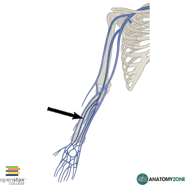
The structure indicated is the radial vein of the forearm.

The venous drainage of the upper limb consists of a superficial system and a deep system. The superficial system drains to the deep system. The veins of the deep system accompany the arteries, and are known as venae comitantes which is the Latin for “accompanying veins” (vena comitans = singular). The veins which accompany the arteries are often in pairs, and are therefore frequently referred to in the plural form – venae comitantes. The radial veins are paired veins which accompany the radial artery in the forearm. The radial veins originate from the deep palmar venous arch and unite with the ulnar veins to drain into the paired brachial veins.

Learn more about the anatomy of the veins of the body in this tutorial.

Viewing all 279 articles
Browse latest View live