Quantcast
Channel: AnatomyZone
Viewing all 279 articles
Browse latest View live

Interspinales

$
0
0

Interspinales

Interspinales

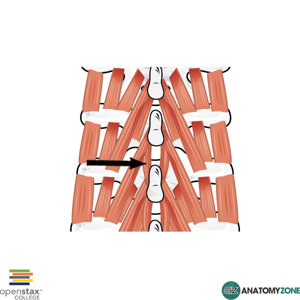
The structure indicated is the interspinales muscle.

The interspinales muscles belong to the deep group of back muscles. The muscles of the back can be considered in terms of a superficial group, an intermediate group and a deep group of muscles. The deep group of back muscles consists of:

  • Spinotransversales muscles
  • Erector spinae muscles and transversospinales muscles
  • Segmental muscles
  • Suboccipital muscles

The interspinales muscles belong to the segmental muscle group of the deep muscles. As the name suggests, the interspinales run between adjacent spinous processes. There is one interspinales muscle on either side, lateral to the interspinous ligament. The interspinales muscles are found in the cervical, thoracic and lumbar region.

Learn about the anatomy of the deep back muscles in this tutorial.


Serratus Anterior

$
0
0

Serratus Anterior

Serratus Anterior

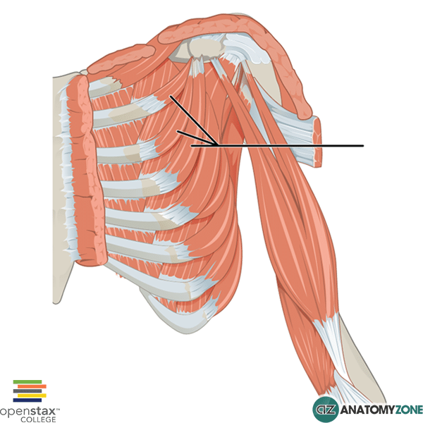
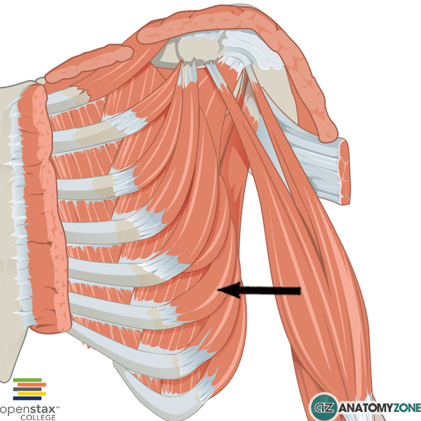
The structure indicated is the serratus anterior muscle.

The serratus anterior muscle originates on the first to eighth ribs laterally on the chest and inserts medially on the scapula. The serratus anterior is divided into three parts:

  1. Serratus anterior superior
  2. Serratus anterior intermediate
  3. Serratus anterior inferior

The serratus anterior is often called the “boxer’s muscle” as it is responsible for protraction of the scapula – a movement which occurs when throwing a punch.

Origin: fleshy slips from lateral parts of 1st to 8th ribs

Insertion: Medial margin of scapula on anterior surface

Innervation: Long thoracic nerve

Action: Protraction and upward rotation of scapula

Parietal Bone

$
0
0

Parietal Bone

Parietal Bone

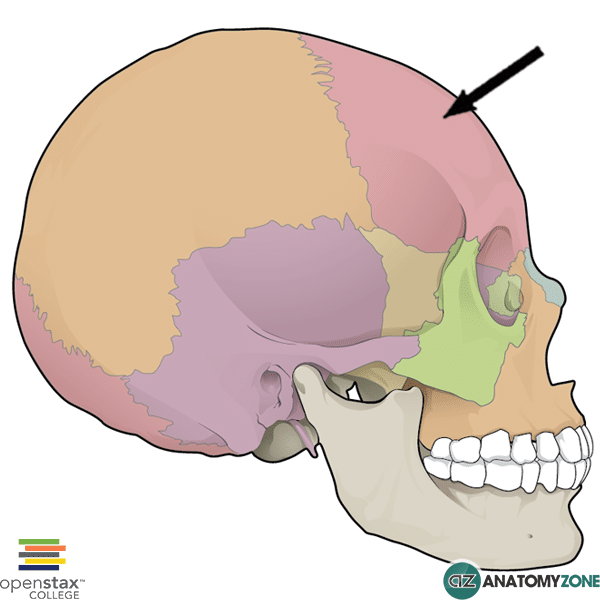
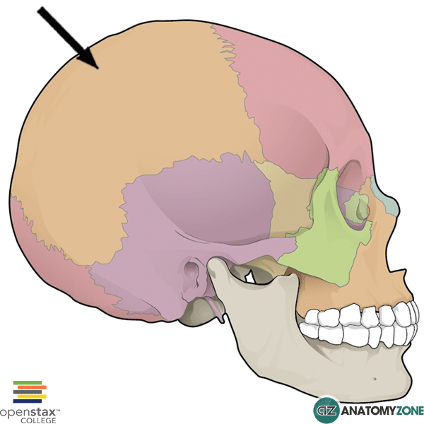
The structure indicated is the parietal bone of the skull.

The parietal bone is one of 8 bones which form the calvaria (the part of the skull which contains the brain). The calvaria consists of:

  • 2 parietal bones
  • 2 temporal bones
  • Frontal bone
  • Sphenoid bone
  • Ethmoid bone
  • Occipital bone

The parietal bone articulates anteriorly with the frontal bone at the coronal suture. Inferiorly it articulates with the temporal bone at the squamous suture. Posteriorly it articulates with the occipital bone via the lamboid suture. It has a small articulation with the sphenoid bone via the sphenoparietal suture at pterion. The parietal bone articulates with the contralateral parietal bone via the sagittal suture.

The parietal bone has the following borders:

  • Sagittal border
  • Frontal border
  • Squamous border
  • Occipital border

The parietal bone has the following angles:

  • Frontal angle
  • Sphenoidal angle
  • Occipital angle
  • Mastoid angle

Learn more about the bones of the skull in this tutorial

Brachioradialis

$
0
0

Brachioradialis

Brachioradialis

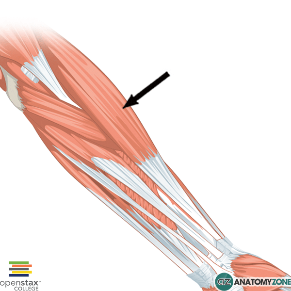
The structure indicated is the brachioradialis muscle of the forearm.

The brachioradalis muscle is one of the muscles of the posterior compartment of the forearm. The posterior compartment consists of a superficial layer of muscles comprised of 7 muscles. The deep layer is comprised of 5 muscles.

Origin: lateral supracondylar ridge of humerus

Insertion: radial styloid process

Innervation: radial nerve

Action: flexes the forearm at the elbow – strongest when forearm is midway between pronation and supination.

Learn more about the muscles of the forearm in this tutorial.

Xiphoid Process

$
0
0

Xiphoid Process

Xiphoid Process

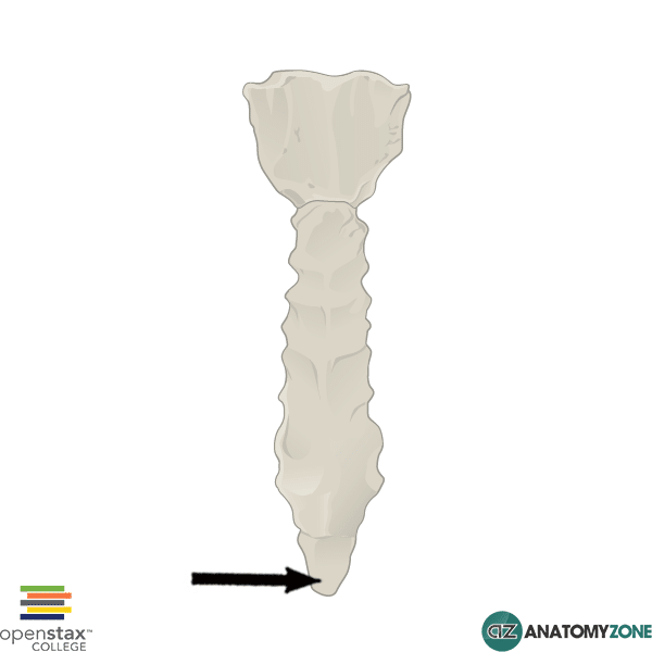
The highlighted structure is the xiphoid process of the sternum.

The sternum consists of three parts:

  1. Manubrium of the sternum
  2. Body of sternum
  3. Xiphoid process

The xiphoid process is the most inferior part of the sternum and is the smallest part.

Learn more about the sternum in this tutorial!

Flexor Digitorum Brevis

$
0
0

Flexor Digitorum Brevis

Flexor Digitorum Brevis

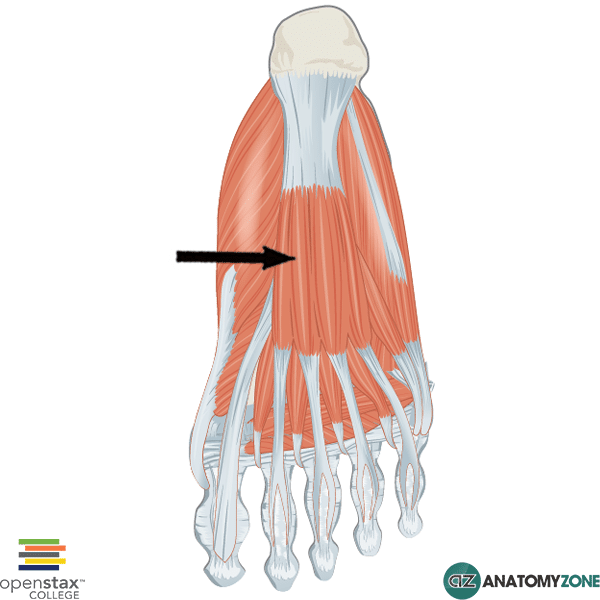
The structure indicated is the flexor digitorum brevis muscle of the foot.

The intrinsic muscles on the plantar aspect of the foot are divided into four layers. The flexor digitorum brevis muscles is found in the first layer of muscles. There are three muscles in the first layer:

  1. Abductor hallucis
  2. Flexor digitorum brevis
  3. Abductor digiti minimi

The flexor digitorum brevis muscle lies above the  central part of the plantar aponeurosis and just below the tendons of flexor digitorum longus.

Origin: medial process of calcaneal tuberosity and plantar aponeurosis.

Insertion: Plantar sides of middle phalanges of lateral four toes

Action: flexes lateral four toes at proximal interphalangeal joint

Innervation: medial plantar nerve from tibial nerve.

Learn more about the muscles of the foot in this tutorial.

Anconeus

$
0
0

Anconeus

Anconeus

The structure indicated is the anconeus muscle of the forearm.

The anconeus muscle is one of the muscles of the posterior compartment of the forearm. The posterior compartment consists of a superficial layer of muscles comprised of 7 muscles. The deep layer is comprised of 5 muscles.

Origin: lateral epicondyle of humerus

Insertion: olecranon, proximal surface of ulna

Innervation: Radial nerve

Action: assists elbow extension, stabilises the elbow joint, abducts ulna during pronation

Learn about the muscles of the forearm in this tutorial.

Thigh Arteries

$
0
0

Hi this is Peter from AnatomyZone, and in this tutorial we’re going to take a look at the arterial supply to the thigh. There are three arteries which supply the thigh, so you’ve got the femoral artery, the obturator artery and the inferior gluteal artery.

Read More

The obturator artery and the inferior gluteal artery are branches of the internal iliac artery, and I looked at these in the previous tutorial where we looked at the branches of the internal iliac.


The femoral artery is the largest branch which enters the thigh and supplies most of the lower limb. In this tutorial we’re going to focus on the various different branches of the femoral artery.


Looking at this model here we can see the external iliac artery and what happens is that it passes under the inguinal ligament to become the common femoral artery. The common femoral artery enters the femoral triangle and it gives off four branches. Now I’ve just zoomed in a little bit further and you can see these four branches here. The first one is the superficial circumflex branch. This one over here is the superficial epigastric branch, and then we’ve got the superficial external pudendal and the deep external pudendal arteries. So those are the four branches which come off the common femoral artery.


Now moving a bit further down we can see that there are several branches which are given off in the thigh. So you’ve got the deep artery of the thigh which is called the profunda femoris artery and you can see that here. This artery is the main supply to the adductor, the extensor and the flexor muscles of the thigh.


The profunda femoris gives off three main branches. You’ve got the lateral circumflex, the medial circumflex and then you’ve got perforating arteries – perforating branches. The lateral circumflex artery has three branches. The first branch you can see here is the descending branch and then coming up to the shaft of the femur, you’ve got two branches. One ascends up onto the neck of the femur. And the other one is called the transverse branch and it winds round laterally, around the proximal shaft of the femur as you can see here.


The medial circumflex artery you can see here coming off a bit more posteriorly from the profunda femoris artery. If I just rotate the model around you can see how it has two branches. It’s got one branch which, like the branch of the lateral circumflex artery, ascends. So there’s an ascending branch which anastomoses with the ascending branch of the the lateral circumflex artery. And there’s also a transverse branch which also anastomoses with the transverse branch of the lateral circumflex artery. So the lateral and medial circumflex branches arise quite proximally on the profunda femoris artery, and if we move distally, we can take a look at the perforating branches.


If I just rotate the model slightly, you can see that there’s three perforating branches and one terminal branch. And these perforating branches penetrate the adductor magnus muscle. And if I rotate around to a posterior view, so what you can see is that these these perforating arteries give off these ascending and descending branches, which unite to form a channel which runs longitudinally.


So we’ve taken a look now at the branches of the common femoral artery and the branches of the deep femoral artery. So what happens after the deep branch is given off, you can see here that it continues down, and the continuation of the common femoral artery after the deep branch is given off, is the superficial femoral artery. So I’m just going to continue to bring the model inferiorly, and you can see what happens as it reaches the distal femur.


The superficial femoral artery then passes through this hole in the adductor magnus muscle, known as the adductor hiatus, and when it does this it becomes the popliteal artery as it enters the posterior compartment.


Now just before the superficial femoral artery leaves via the adductor hiatus, it gives off this branch known as the descending genicular artery. The descending genicular artery itself has two main branches, you’ve got the saphenous branch which passes medially and then anastomoses with the medial superior genicular – which we’ll take a look at in the next part of the tutorial series. And the other branch is this articular branch, so you’ve got lots of articular branches which anastomose around the knee joint.


So that’s a look at some of the branches which provide the arterial supply to the thigh. In the next tutorials we’ll take a look at the arterial supply to the knee, the leg, and the foot.
Pectoralis Minor

$
0
0

Pectoralis Minor

Pectoralis Minor

The structure indicated is the pectoralis minor muscle.

The pectoralis minor muscle lies deep to the much larger, powerful pectoralis major muscle.

The pectoralis minor muscle is covered anteriorly by the clavipectoral fascia. This muscle divides the axillary artery into three parts. Part 1 lies proximally to the pectoralis minor, part 2 is the part of the axillary artery covered by the pectoralis minor, and the third part lies distal to the pectoralis minor.

The pectoralis minor inserts onto the coracoid process of the scapula. There are three muscles which attach to the coracoid process:

  1. Short head of biceps brachii
  2. Coracobrachialis
  3. Pectoralis minor

Origin: anteromedial surfaces of ribs 3,4,5

Insertion: coracoid process of scapula

Action: depresses shoulder, protracts scapula

Innervation: medial pectoral nerve

Inferior Vena Cava

$
0
0

Inferior Vena Cava

Inferior Vena Cava

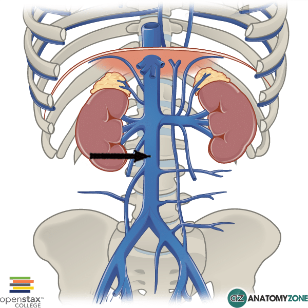
The structure indicated is the inferior vena cava.

The inferior vena cava is responsible for carrying deoxygenated blood into the right atrium of the heart from the lower part of the body. The inferior vena cava is formed at the point of union of the right and left common iliac veins at the L5 vertebral level.

Several veins drain into the inferior vena cava:

  • Lumbar veins
  • Right gonadal vein (the left gonadal vein drains into the left renal vein)
  • Renal veins
  • Right suprarenal vein
  • Inferior phrenic vein
  • Hepatic veins

The inferior vena cava ascends through the abdomen immediately anterior to the vertebral column on the right side, adjacent to the abdominal aorta which sits on the left.

Learn more about the veins of the body in this tutorial.

Aortic Arch

$
0
0

Aortic Arch

Aortic Arch

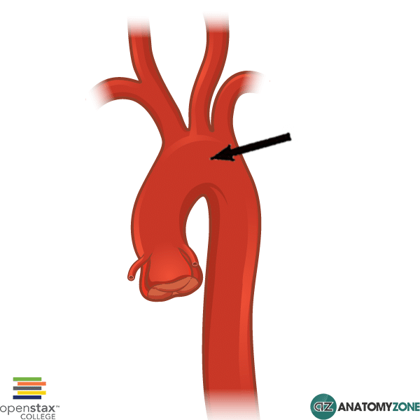
The structure indicated is the arch of the aorta.

The aorta arises from the left ventricle of the heart and supplies the body with oxygenated blood. The aorta can be divided into thoracic and abdominal parts (the thoracic aorta pierces the diaphragm to enter the abdominal cavity, becoming the abdominal aorta). The thoracic aorta consists of:

  • Ascending aorta
  • Aortic arch
  • Descending (thoracic) aorta

The aortic arch lies in the superior mediastinum and begins and ends at the level of the angle of Louis (sternal angle) which lies at the level of the intervertebral disc between T4/T5.

The arch of the aorta gives rise to three branches:

  1. Brachiocephalic trunk
  2. Left common carotid
  3. Left subclavian artery

You can remember the branches of the aortic arch using the mnemonic ABC’S – Aorta – Brachiocephalic trunk, Common carotid (left), Subclavian (left).

Learn more about the great vessels in this tutorial

Psoas Major

$
0
0

Psoas Major

Psoas Major

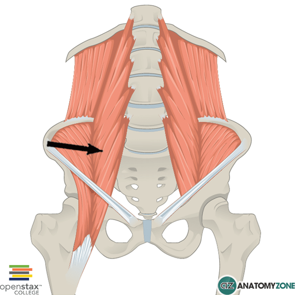
The structure indicated is the psoas major muscle

The psoas major muscle is one of the muscles which makes up the posterior abdominal wall.

There are four muscles which contribute to the posterior abdominal wall:

  1. Psoas major
  2. Psoas minor
  3. Quadratus lumborum
  4. Iliacus

The iliacus muscle combines with the psoas major muscle to enter the anterior compartment of the thigh and insert via a common tendon on the lesser trochanter of the femur. The iliacus and the psoas major muscles are thus collectively referred to as the iliopsoas muscle – they act synergistically to flex the thigh at the hip joint, as well as acting to cause lateral rotation of the thigh. The iliopsoas muscles are the strongest hip flexors.

Origin: Transverse processes of L1-L5, vertebral bodies and discs of T12-L5

Insertion: Lesser trochanter of femur

Action: hip flexion, lateral rotation of thigh

Innervation: fibres of L1-L3 of lumbar plexus

Learn more about the muscles of the posterior abdominal wall in this tutorial.

Sternal Angle

$
0
0

Sternal Angle

Sternal Angle

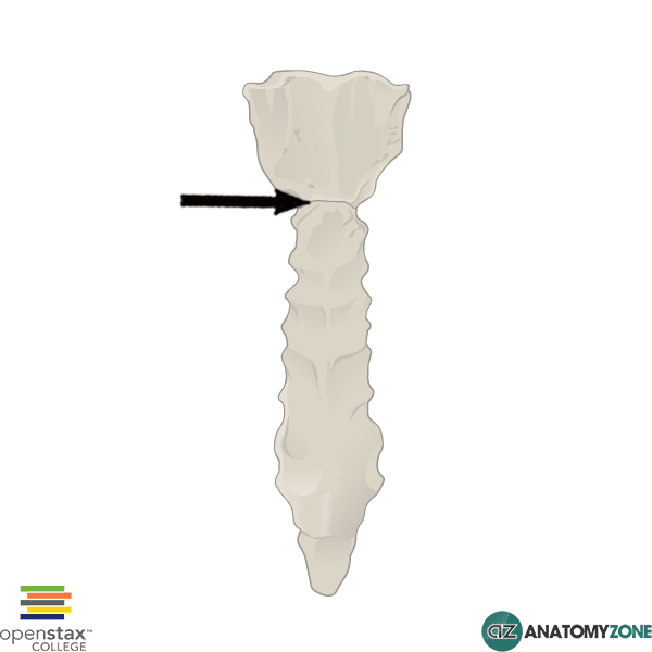
The diagram indicates the sternal angle of Louis.

The sternal angle is the angle formed between the manubrium of the sternum and the body of the sternum (manubriosternal junction), and is an important anatomical landmark. It marks the level of the 2nd pair of costal cartilages which lies at the level of the intervertebral disc between thoracic vertebrae 4 and 5. This level represents several important anatomical features:

  • Beginning and end of the aortic arch
  • Bifurcation of the trachea
  • Bifurcation of the pulmonary trunk
  • Left recurrent laryngeal loops under arch of aorta
  • Ligamentum arteriosum lies at this level
  • Azygos vein drains into superior vena cava
  • Cardiac plexus
  • Thoracic duct empties into left subclavian vein

A helpful mnemonic for remembering the structures that lies at the level of the sternal angle is RATPLANT:

Rib (2nd), Aortic arch, Trachea birfurcation, Pulmonary trunk bifurcation, Left recurrent laryngeal (and Ligamentum arteriosum), Azygos vein, Nerves – cardiac plexus, Thoracic duct

Learn about the sternal angle in this tutorial.

Sinoatrial Node

$
0
0

Sinoatrial Node

Sinoatrial Node

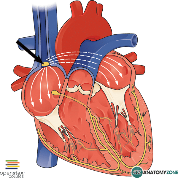
The structure indicated is the sinoatrial node

The conduction system of the heart is responsible for the coordinated contraction of the atria and ventricles and is comprised of the following main structures:

  • Sinoatrial node
  • Atrioventricular node
  • Atrioventricular bundle of His
  • Right and left bundle branches
  • Purkinje fibres

The sinoatrial node (sinoatrial node), often referred to as the SA node is the pacemaker of the heart located in the right atrium and is responsible for generating normal sinus rhythm.

The sinoatrial node is located lateral to the junction where the superior vena cava enters the right atrium. Electrical signals generated from the sinoatrial node spread across the atria to the atrioventricular node, which is located close to the opening of the coronary sinus in the right atrium.

It receives parasympathetic (cranial nerve X – vagus nerve) and sympathetic innervation (T1-4 spinal nerves). Parasympathetic activation decreases sinoatrial firing and thus decreases heart rate. Sympathetic activation does the opposite, causing increase sinoatrial firing and increases heart rate.

Frontal Bone

$
0
0

Frontal Bone

Frontal Bone

The structure indicated is the frontal bone of the skull

The frontal bone is one of 8 bones which form the calvaria (the part of the skull which contains the brain). The calvaria consists of:

  • 2 parietal bones
  • 2 temporal bones
  • Frontal bone
  • Sphenoid bone
  • Ethmoid bone
  • Occipital bone

The frontal bone consists of three parts:

  1. Squamous part
  2. Orbital part
  3. Nasal part

The frontal bone articulates with the following bones:

  • Sphenoid bones
  • Ethmoid bone
  • Parietal bones
  • Nasal bones
  • Maxillae bones
  • Lacrimal bones
  • Zygomatic bones

The point at which the frontal bones meet the parietal bones in the midline is called bregma – this is where the coronal suture is intersected by the sagittal suture.

The point at which the frontal bones meet the two nasal bones is known as nasion.

Learn more about the bones of the skull in this tutorial.


Knee – Arterial Supply

$
0
0

Hi this is Peter from AnatomyZone, and in this tutorial we’re going to take a look at the arterial supply to the knee. So this follows on from the previous tutorial on the arterial supply to the thigh.

Read More

The arteries which supply the knee are derived from branches of the femoral and popliteal arteries. You’ve got the descending branch of the lateral circumflex artery, the descending genicular branch of the superficial femoral artery, you’ve got genicular arteries derived from the popliteal artery, recurrent branches from the tibial arteries and you’ve also got the circumflex fibular branch. These arteries all contribute to this rich anastomotic network around the knee.


So if I just rotate the model around to a posterior view, we can see the popliteal artery. The popliteal artery is a continuation of the SFA – the superficial femoral artery as it passes through the adductor hiatus. The popliteal artery then terminates by dividing into the anterior and posterior tibial arteries. So you’ve got the anterior tibial artery here in yellow and the posterior tibial artery here in green.


I’ve just zoomed in a little bit closer and we’re now looking at the genicular arteries, which are branching off the popliteal artery. So as you can see, you’ve got a superior and inferior set of genicular arteries. And you’ve got genicular arteries on either side, so you’ve got medial and lateral genicular arteries. At the top we’ve got the lateral superior genicular and the medial superior genicular, and at the bottom we’ve got the lateral inferior genicular, and the medial inferior genicular.


If I just rotate the model around anteriorly, you can see how these arteries form this network of vessels around the knee. All these genicular arteries anastomose. So just rotating the model around to a medial view of the knee, we can see the medial superior and the medial inferior genicular arteries. And you can see that they receive this anastomosing branch from the descending genicular artery, which is a branch from the superficial femoral artery.


And then rotating around to a lateral view, taking a look at the lateral superior and the lateral inferior genicular arteries, we can see that the lateral inferior genicular artery receives two branches. So in this model you can see anteriorly, it receives this branch which comes off the anterior tibial artery, known as the anterior tibial recurrent artery. And if I rotate the model slightly posteriorly, you can see this branch arising from the posterior tibial artery and winding around the fibula, and then anastomosing with the lateral inferior genicular artery. This is the circumflex fibular artery.


At the midpoint of the popliteal artery entering into the posterior aspect of the knee joint, there’s another genicular artery known as the middle genicular artery. If I rotate the model around slightly, you can see this branch highlighted in green, entering into the posterior aspect of the knee joint. The middle genicular artery pierces the oblique popliteal ligaments to supply the synovial membrane and the cruciate ligaments of the knee.


Now also on this model, visible from this view are the sural arteries. The sural arteries are superficial arteries arising from the popliteal artery, and you’ve got a medial and lateral sural artery, and these supply the gastrocnemius, the soleus and the plantaris muscles. In blue, we have the medial sural artery, and in purple we have the lateral sural artery.


So we’ve taken a look now at various different branches given off from the arteries in the thigh and the leg to supply the knee joint. In the next tutorial we’ll take a look at the branches of the arteries in the leg.

Iliopsoas Muscle

$
0
0

Iliopsoas Muscle

Iliopsoas Muscle

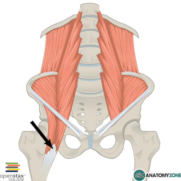
The structure indicated is the iliopsoas muscle.

The iliopsoas muscle collectively refers to the distal ends of the psoas major and iliacus muscles which exit the pelvis to enter the anterior compartment of the thigh and insert onto the lesser trochanter of the femur.

The iliopsoas muscle is the strongest hip flexor.

Origin: Iliac fossa and vertebral bodies of T12 to L5

Insertion: Lesser trochanter of femur

Innervation: anterior rami of lumbar plexus (L2-L4)

Action: Hip flexion, lateral rotation of femur

Learn more about the muscles of the anterior compartment of the thigh in this tutorial.

Occiptofrontalis

$
0
0

Occipitofrontalis

Occipitofrontalis - Frontal Belly

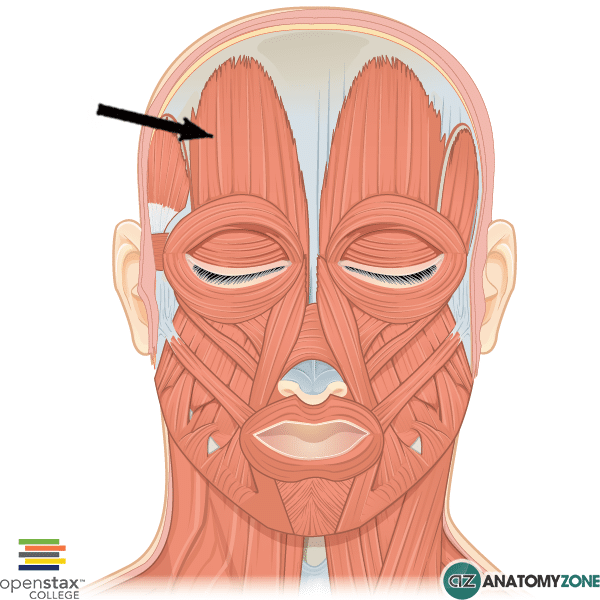
The structure indicated is the frontal belly of the occipitofrontalis muscle.

The occipitofrontalis muscle is involved in facial expression and is comprised of two muscle bellies:

  1. Frontal belly
  2. Occipital belly

Contraction of the frontal belly causes wrinkling of the forehead and raising of the eyebrows. Contraction of the occipital belly pulls the scalp back posteriorly. As a muscle of facial expression, the occipitofrontalis muscle is innervated by the facial nerve (cranial nerve VII).

In between the frontal and occipital bellies, is an aponeurotic tendon which connects two bellies.

Learn more about the muscles of facial expression in this tutorial.

Nasal Bone

$
0
0

Nasal Bone

Nasal Bone

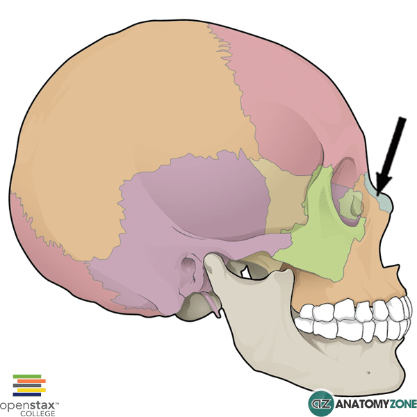
The structure indicated is the nasal bone of the facial skeleton.

The skull consists of the calvaria (the part which contains the brain), and the facial skeleton (viscerocranium) which forms the anterior and inferior part of the skull.

The facial skeleton consists of the following bones:

  • Nasal bones
  • Lacrimal bones
  • Palatine bones
  • Zygomatic bones
  • Maxillae bones
  • Inferior nasal conchae
  • Vomer

There are two paired nasal bones. The nasal bone has four articulations:

  1. Frontal bone (superiorly)
  2. Ethmoid bone (posteriorly)
  3. Maxillae (laterally)
  4. Nasal bone (medially)

Learn more about the anatomy of the facial skeleton in this tutorial.

Atrioventricular Node

$
0
0

Atrioventricular Node

Atrioventricular Node

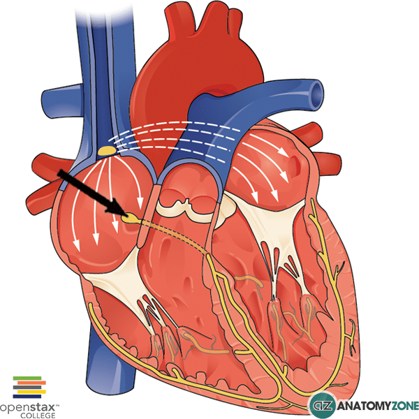
The structure indicated is the atrioventricular node.

The conduction system of the heart is responsible for the synchronised contraction of the atria and ventricles and is comprised of the following main structures:

  • Sinoatrial node
  • Atrioventricular node
  • Atrioventricular bundle of His
  • Right and left bundle branches
  • Purkinje fibres

The atrioventricular node is located centrally in the Triangle of Koch, between the atria and ventricles in the infero-posterior portion of the interatrial septum close to the opening of the coronary sinus. The triangle of Koch has the following boundaries:

  1. Ostium of coronary sinus
  2. Tendon of Todaro
  3. Anterior-septal leaflet commissure

The atrioventricular nodes receives signals from the sinoatrial node. The impulses from the atrioventricular node then pass to the Bundle of His and then follow the left and right bundle branches through the interventricular septum and ultimately to the Purkinje fibres which propagate the signal into the ventricles.

The atrioventricular node delays the impulse generated by the atria, which allows the atria time to contract and propel blood into the ventricles before ventricular contraction.

Viewing all 279 articles
Browse latest View live