Quantcast
Channel: AnatomyZone
Viewing all 279 articles
Browse latest View live

Squamous Suture

$
0
0

Squamous Suture

Squamous Suture

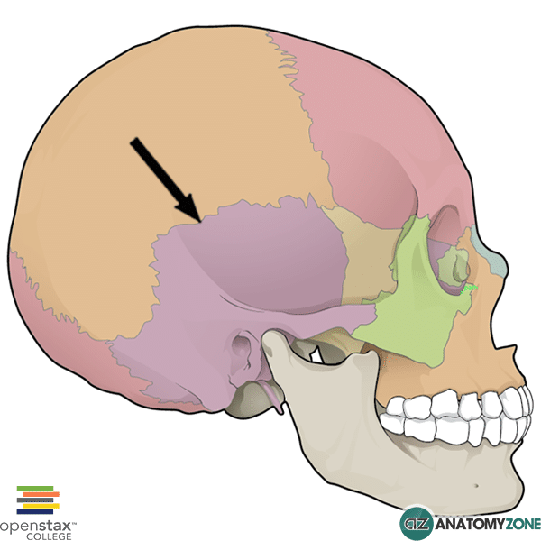
The structure indicated is the squamous suture of the skull.

Sutures are junctions between the bones of the skull – they are a type of fibrous joint, bound together by Sharpey’s fibres.

The squamous suture forms the joint of the skull between the squamous part of the temporal bone and the inferior aspect of the parietal bone. Posteriorly, the squamous suture becomes the parietomastoid suture where the mastoid process articulates with the parietal bone. Anteriorly, the squamous suture extends towards pterion.

Learn more about the sutures of the skull in this tutorial.


Left Subclavian Artery

$
0
0

Left Subclavian Artery

Left Subclavian Artery

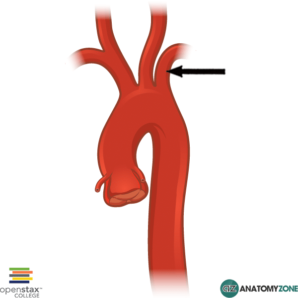
The structure indicated is the left subclavian artery.

The subclavian arteries are located under (“sub”) the clavicle (“clavian”) and receive blood from the aortic arch. The subclavian arteries supply the arms, with some branches that extend to supply the head.

The branches of the aortic arch are:

  • Brachiocephalic artery
  • Left common carotid artery
  • Left subclavian artery

The left subclavian artery comes directly off the arch of the aorta, whereas on the right side of the body, the brachiocephalic artery splits, giving rise to the right subclavian artery, and the right common carotid artery.

The subclavian artery becomes the axillary artery at the lateral border of the first rib. It can be thought of in three parts, and gives rise to several branches:

  • First part: vertebral artery, internal thoracic artery, thyrocervical trunk
  • Second part: costocervical trunk
  • Third part: dorsal scapular artery

Mandibular Notch

$
0
0

Mandibular Notch

Mandibular Notch

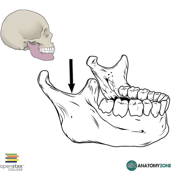
The structure indicated is the mandibular notch.

The ramus of the mandible forms the lateral portion of the mandible. It has two processes:

  1. Coronoid process
  2. Condyloid process

These processes extend superiorly from the ramus of the mandible. The coronoid process is located anteriorly, and the condyloid process is located posteriorly. In between these two processes is a concave notch, known as the mandibular notch.

Passing through this notch are the following structures:

  • Masseteric nerve
  • Masseteric artery
  • Masseteric vein

Learn more about the anatomy of the mandible in this tutorial.

Abductor Digiti Minimi

$
0
0

Abductor Digiti Minimi

Abductor Digiti Minimi

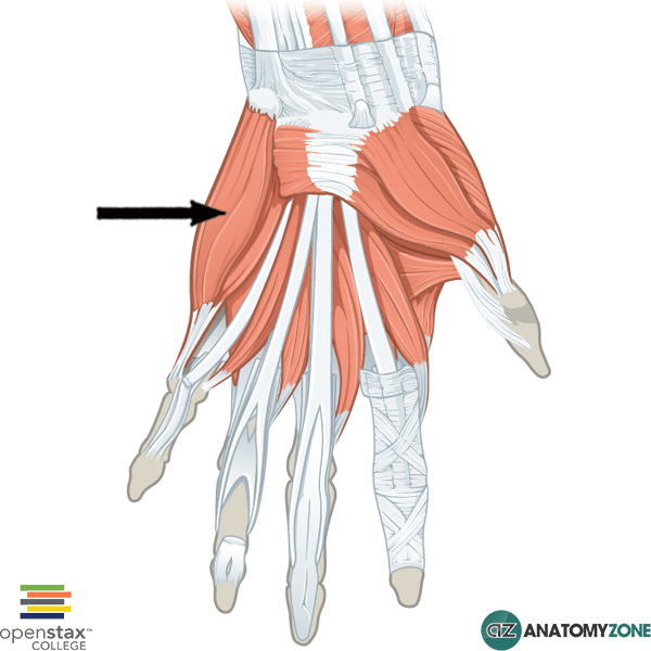
The structure indicated is the abductor digiti minimi muscle of the hand.

The abductor digiti minimi muscle is one of the intrinsic muscles of the hand that belongs to the hypothenar group.

The hypothenar group of intrinsic hand muscles consist of three muscles:

  1. Abductor digiti minimi
  2. Opponens digiti minimi
  3. Flexor digit minimi brevis

All the intrinsic muscles of the hand, except the thenar muscles and the lateral two lumbrical muscles are innervated by the deep branch of the ulnar nerve. The thenar muscles and the lateral two lumbrical muscles are innervated by the median nerve.  A useful mnemonic for remembering this is MEATLOAF. “MEAT” refers to the Median nerve, and LOAF refers to the muscles which it innervates: Lateral two lumbricals, Opponens pollicis, Abductor pollicis brevis, Flexor pollicis brevis.

Origin: Pisiform

Insertion: Medial base of proximal phalanx of little finger

Innervation: Deep branch of ulnar nerve

Action: Abducts little finger

Learn all about the anatomy of the muscles of the hand in this tutorial.

Perpendicular Plate of Ethmoid Bone

$
0
0

Perpendicular Plate of Ethmoid Bone

Perpendicular Plate of Ethmoid Bone

The structure indicated is the perpendicular plate of the ethmoid bone.

The ethmoid bone is one of the bones in the skull that separates the nasal cavity from the brain. There are three parts of which the ethmoid bone is comprised:

  1. Cribriform plate
  2. Ethmoidal labyrinth (one on either side)
  3. Perpendicular plate

The perpendicular plate of the ethmoid bone extends inferiorly from the base of the cribriform plate and contributes to the median nasal septum. It has the following articulations:

  • Anteriorly: spine of frontal bone
  • Posteriorly: sphenoidal crest
  • Inferiorly: septal cartilage anteriorly, vomer posteriorly

Learn more about the bones of the skull in this tutorial

Anterior Tibial Artery

$
0
0

Anterior Tibial Artery

Anterior Tibial Artery

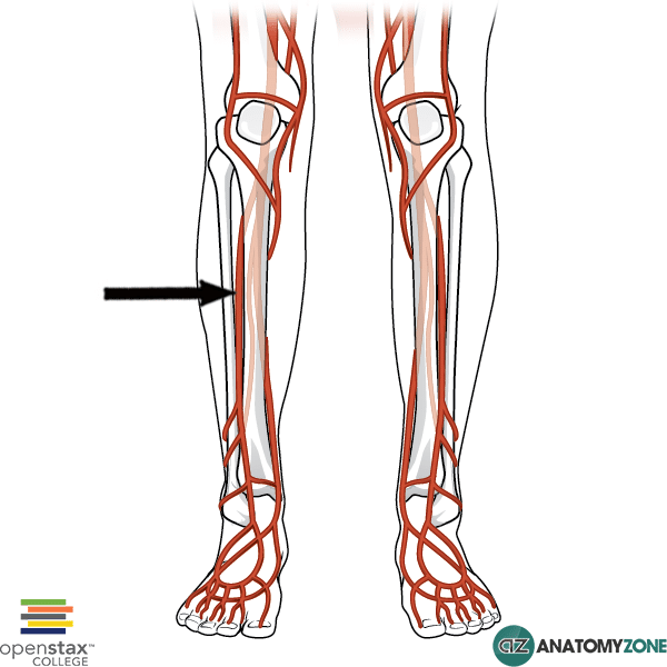
The structure indicated is the anterior tibial artery of the leg.

The anterior tibial artery is a branch of the popliteal artery, which supplies the anterior compartment of the leg. The popliteal artery arises from the superficial femoral artery as it passes from the anterior compartment of the thigh to the posterior compartment via the adductor hiatus – an opening in the distal adductor magnus muscle. The popliteal artery gives rise to two branches, the anterior tibial artery and the posterior tibial artery. The anterior tibial artery passes through an opening superiorly in the interosseous membrane between the tibia and the fibula.

The anterior tibial artery descends the length of the leg, accompanied by the tibial vein, and becomes the dorsal pedis artery on the dorsal surface of the foot.

The anterior tibial artery has the following branches:

  • Anterior tibial recurrent artery
  • Posterior tibial recurrent artery
  • Anterior medial malleolar artery
  • Anterior lateral malleolar artery
  • Muscular branches
  • Perforating branches

Adductor Magnus

$
0
0

Adductor Magnus

Adductor Magnus Muscle

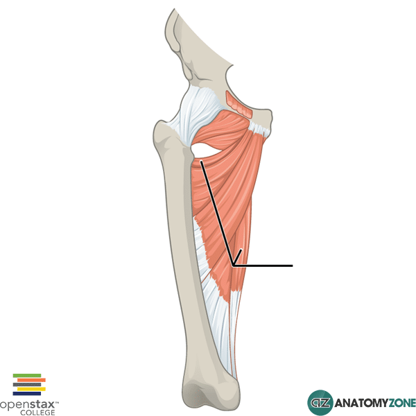
The structure indicated is the adductor magnus muscle of the thigh.

The adductor magnus muscle is one of six muscles in the medial compartment of the thigh. The medial compartments consists of the following muscles:

  • Gracilis
  • Pectineus
  • Adductor longus
  • Adductor brevis
  • Adductor magnus
  • Obturator externus

The adductor magnus muscle is the largest muscle in the medial compartment. It lies deep to the adductor brevis and the adductor longus. It has two parts to it:

  1. Adductor part
  2. Hamstring part

The adductor part originates on the ischiopubic ramus, and as the name suggests, contraction causes adduction, as well as medial rotation of the thigh.

The hamstring part originates on the ischial tuberosity and contraction results in extension of the hip.

Distally, there is a hole in the adductor magnus muscle known as the adductor hiatus (not shown in image). This allows the femoral artery and veins to pass from the anterior compartment of the thigh into the posterior compartment.

Origin: adductor part – anteroinferior pubic ramus; hamstring part – ischial tuberosity

Insertion: adductor part – linea aspera, hamstring part – adductor tubercle

Action: adduction of hip (adductor part), extension of hip (hamstring part)

Innervation: adductor part – obturator nerve, hamstring part – tibial nerve (from sciatic nerve)

Learn all about the anatomy of the thigh muscles in this tutorial.

Lateral Cuneiform Bone

$
0
0

Lateral Cuneiform Bone

Lateral Cuneiform Bone

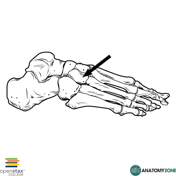
The structure indicated is the lateral cuneiform bone of the foot.

The foot contains three groups of bones:

  1. Tarsals
  2. Metatarsals
  3. Phalanges

The lateral cuneiform bone is one of seven tarsal bones. There are three groups of tarsal bones:

  • Proximal group
  • Intermediate group
  • Distal group

The lateral cuneiform bone belongs to the distal group of tarsal bones. There are three cuneiform bones:

  • Lateral cuneiform
  • Intermediate cuneiform
  • Medial cuneiform

Anteriorly they articulate with the medial three metatarsals. Posteriorly they articulate with the navicular bone, and laterally they articulate with the cuboid bone.

Learn more about the bones of the foot in this anatomy tutorial.


Sacrospinous Ligament

$
0
0

Sacrospinous Ligament

Sacrospinous Ligament

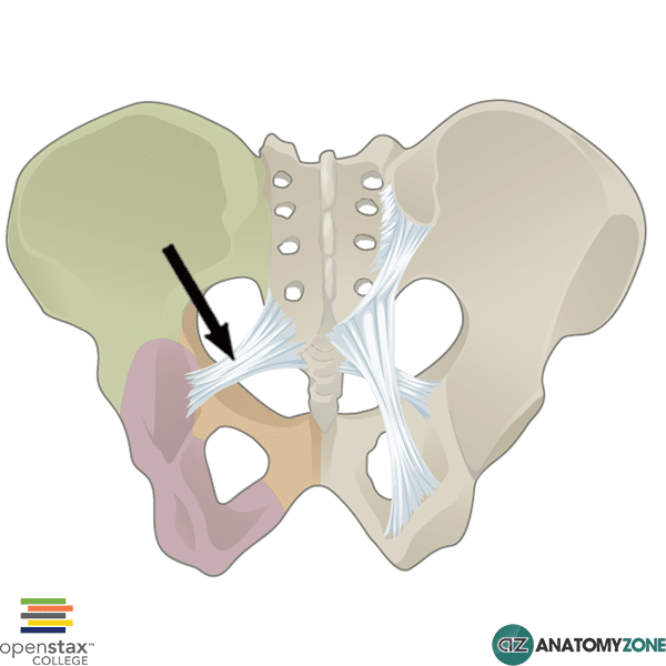
The structure indicated is the sacrospinous ligament.

The sacrospinous ligament attaches at one end to the ischial spine, and at the other end to the lateral aspect of the sacrum and coccyx, lying deep to the sacrotuberous ligament. The sacrotuberous ligament runs from the sacrum to the ischial tuberosity.

The sacrotuberous ligament converts the greater sciatic notch into the greater sciatic foramen. Likewise, it converts the lesser sciatic notch into the lesser sciatic foramen.

Learn more about the bones of the pelvis in this tutorial.

Middle Nasal Concha

$
0
0

Middle Nasal Concha

Middle Nasal Concha

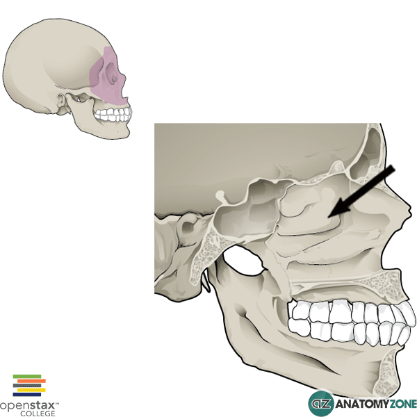
The structure indicated is the middle nasal concha.

There are three nasal conchae which make up the lateral wall of the nasal cavity. The nasal conchae are curved bones which create channels for the flow of air through the nose. There are four air channels:

  • Inferior nasal meatus (underneath inferior nasal concha)
  • Middle nasal meatus (underneath medial nasal concha)
  • Superior nasal meatus (underneath superior nasal concha)
  • Spheno-ethmoidal recess (between superior nasal concha and nasal roof)

The superior and middle nasal concha are formed from the ethmoidal labyrinths of the ethmoid bone. The ethmoidal labyrinths lie on either side of the central cribriform plate, and consist of two sheets of bone (medial and lateral). The superior and middle nasal conchae are formed from the medial sheet of bone.

The inferior nasal concha on the other hand is formed from the maxillae bones of the facial skeleton. Thus, the inferior nasal conchae is considered a facial bone, whereas the middle and superior nasal conchae are considered cranial bones.

Learn more about the anatomy of the skull in this tutorial.

Internal Jugular Vein

$
0
0

Internal Jugular Vein

Internal Jugular Vein

The structure indicated is the internal jugular vein.

The jugular veins are responsible for returning deoxygenated blood from the head to the heart via the superior vena cava. There are two jugular veins:

  • External jugular vein
  • Internal jugular vein

The external jugular vein drains into the subclavian vein. The internal jugular vein joins the subclavian vein to form the brachiocephalic vein. The brachiocephalic veins (innominate vein) on either side then join one another to form the superior vena cava, to bring blood into the right atrium of the heart. The external jugular vein runs superficial to the sternocleidomastoid muscle, the internal jugular vein on the other hand runs deep to this muscle inside the carotid sheath along with the common carotid artery and the vagus nerve.

Due to the direct pathway between the internal jugular vein and the right atrium of the heart (there is only a single pair of valves between the two), the internal jugular vein can be used as a “barometer” to measure the pressure in the venous system due to problems arising in the heart and lungs, resulting in problems with the right side of the heart. The jugular venous pressure (JVP) is the pulsation seen in the internal jugular vein, allowing one to estimate the pressure in the right atrium. It may be raised in conditions such as right ventricular failure.

Soleal Line

$
0
0

Soleal Line

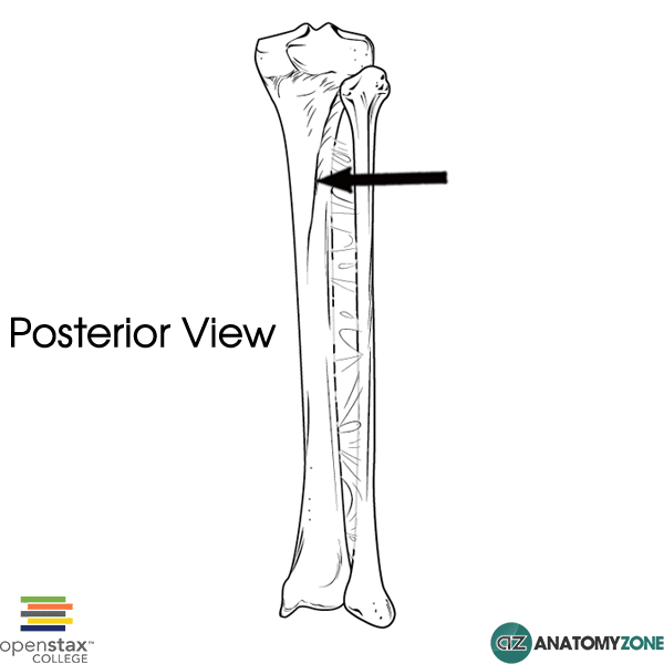
Soleal Line of Tibia

The structure indicated is the soleal line of the tibia.

The soleal line is an oblique line visible on the posterior surface of the tibia. The popliteus muscle inserts above the soleal line.

Three other muscles in the posterior compartment of the leg take their origin, or part of their origin from the soleal line:

  • Soleus
  • Flexor digitorum longus
  • Tibialis posterior

Learn all about the anatomy of the tibia and the fibula.

Lower Limb Arteries

Hyoglossus Muscle

$
0
0

Hyoglossus Muscle

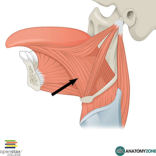
Hyoglossus Muscle

The structure indicated is the hyoglossus muscle of the tongue.

The hyoglossus muscle is one of the extrinsic muscles of the tongue. The muscles involved with the tongue consist of intrinsic muscles which lie within the tongue itself, and the extrinsic muscles which attach to the tongue and are responsible for depression, elevation, protraction and retraction of the tongue.

There are four sets of extrinsic tongue muscles:

  1. Genioglossus
  2. Hyoglossus
  3. Palatoglossus
  4. Styloglossus

Origin: Greater horn and body of hyoid bone

Insertion: Lateral aspect of tongue

Action: Depression and retraction of tongue

Innervation:  Hypoglossal nerve (cranial nerve XII)

Learn all about the anatomy of the cranial nerves and the extrinsic muscles of the tongue and hyoid bone.

Posterior Interventricular (Descending) Artery

$
0
0

Posterior Interventricular (Descending) Artery

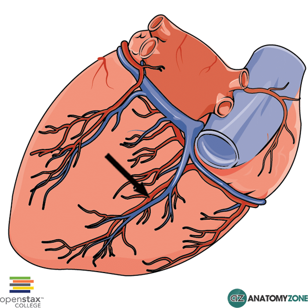
Posterior Interventricular (Descending) Artery

The structure indicated is the posterior interventricular artery (posterior descending artery) of the heart.

The posterior interventricular artery determines the “dominance” of the coronary blood supply to the heart. For example, if the posterior interventricular artery arises from the right coronary artery, the heart is said to be “right coronary dominant”. In the majority of people, the posterior interventricular artery will usually arise from the right coronary artery. If the posterior interventricular artery arises from the left coronary artery, it will branch from the circumflex coronary artery.

Co-dominance occurs when the posterior interventricular artery arises from an anastomosis between the left and right coronary arteries.

The posterior interventricular artery runs in the posterior interventricular sulcus and supplies the posterior third of the interventricular septum (the rest is supplied by the anterior interventricular artery)

Learn more about the blood supply to the heart in this anatomy tutorial.


Blood Capillary

$
0
0

Blood Capillary

Blood Capillary

The diagram illustrates a capillary.

Capillaries are the smallest type of blood vessel in the human body. They are responsible for the transfer of oxygen, carbon dioxide, fluids, nutrients, and waste products to and from the body’s tissues. Capillaries form a connection between arterioles and venules. Organ systems that are highly metabolically active have dense capillary networks, such as the muscular system and renal system.

Structure: Thin walled one cell layer thick endothelium (simple squamous epithelium).

Blood flow sequence

heart –> arteries –> arterioles –> capillaries –> venules –> veins –> heart

There are three types of blood capillaries:

  1. Continuous
  2. Fenestrated
  3. Sinusoid

In some capillaries, the blood flow into the capillary is regulated by precapillary sphincters (bands of smooth muscle) at their point of origin from the arteriole off which it branches. These precapillary sphincters are found in the mesenteric microcirculation.

Great Cardiac Vein

$
0
0

Great Cardiac Vein

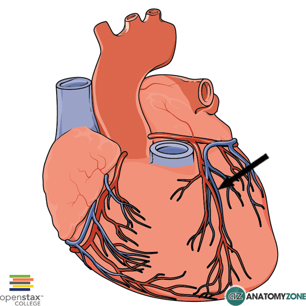
Great Cardiac Vein

The structure indicated is the great cardiac vein.

The coronary sinus lies posteriorly on the heart in the coronary sulcus and receives four cardiac veins:

  • Great cardiac vein
  • Middle cardiac vein
  • Small cardiac vein
  • Posterior cardiac veins

The great cardiac vein lies in the anterior interventricular sulcus together with the anterior interventricular artery (left anterior descending), and for this reason it is sometimes referred to as the anterior interventricular vein.

The great cardiac vein begins at the apex of the heart and ascends in the anterior interventricular sulcus to reach the coronary sulcus, where it then runs along with the circumflex artery to the posterior aspect of the heart to drain into the coronary sinus.

Learn more about the anatomy of the venous drainage of the heart in this tutorial.

Tricuspid Valve

$
0
0

Tricuspid Valve

Tricuspid Valve

The structure indicated is the tricuspid valve of the heart.

There are four sets of valves in the heart. Between the atria and the ventricles are the atrioventricular valves; on the right is the tricuspid valve, on the left is the mitral (bicuspid) valve.

Between the ventricles and the aorta/pulmonary trunk are the semilunar valves. The pulmonary valve is located between the right ventricle and the pulmonary trunk, and the aortic valve is located between the left ventricle and the aorta.

The atrioventricular valves are controlled by papillary muscles, which are specialised structures in the ventricles that attach to the valves via string-like extensions known as chordeae tendineae. They contract to prevent the atrioventricular valves from inverting and allowing blood to flow backwards into the atria.

There are three papillary muscles in the right ventricle which attach to the tricuspid valve:

  1. Anterior
  2. Posterior
  3. Septal

Learn more about the anatomy of the right ventricle in this tutorial.

Splenius Capitis

$
0
0

Splenius Capitis

Splenius Capitis

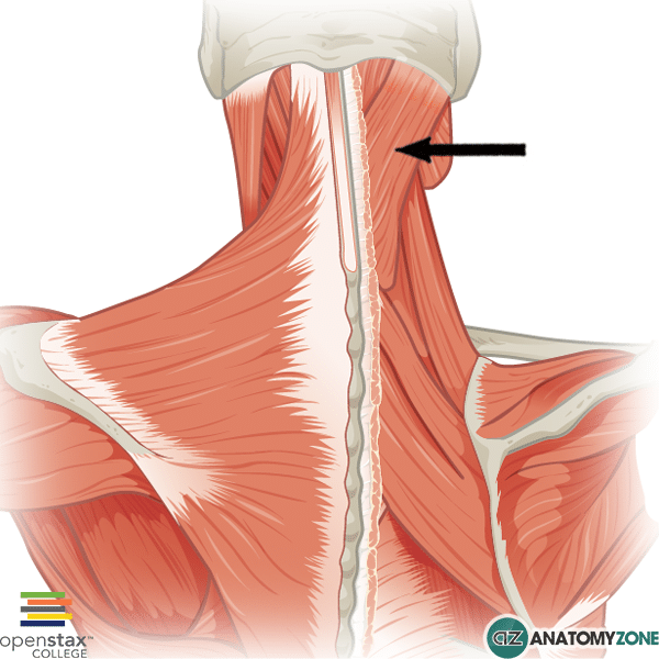
The structure indicated is the splenius capitis muscle.

The splenius capitis muscle is one of 8 muscles in the posterior triangle of the neck.

The posterior triangle of the neck is bounded as follows:

  • Anteriorly: posterior border of sternocleidomastoid
  • Posteriorly: anterior border of trapezius
  • Apex: Occipital bone between attachment sites of trapezius and sternocleidmastoid
  • Base: middle third of clavicle

Origin: Lower half of ligamentum nuchae and spinous processes of C7-T4

Insertion: Mastoid process, and part of skull inferior to lateral superior nuchal line.

Action: Contraction together – extension of the neck. Contraction individually – Lateral flexion of neck and rotation of head to same side.

Innervation: Posterior rami C3-C6

Learn more about the muscles of the posterior triangle of the neck in this anatomy tutorial.

Internal Iliac Artery

$
0
0

Hi this is Peter from AnatomyZone and in this tutorial we’re going to take a look at the internal iliac artery.  In this model, what we’re looking at is the abdominal aorta dividing into the common iliac arteries, which then divide into the external and internal iliac arteries.

Read more

What we’re now looking at is the right internal iliac artery, and we’ve removed the left, and we’ve removed the external iliac arteries. I’ve just zoomed in a little bit closer, and you can see the two trunks. One extends posteriorly and is known as the posterior trunk, and the other one, which I’ve removed the branches from and we’ll come on to talk about in a moment, is the anterior trunk.   you’ve got the anterior and posterior trunk.


The posterior trunk has three branches, you’ve got the iliolumbar artery, the lateral sacral arteries and the superior gluteal artery.  I’ve just zoomed out a little bit, and we’ll take a look at the iliolumbar artery.   This is this artery here. If I just rotate the model around a bit further, you can see the two branches of the iliolumbar artery. You’ve got the iliac branch and the lumbar branch. I’ve just zoomed in for a better view and you can see the lumbar branch which extends superiorly just adjacent to the lumbar vertebrae. And then taking a look at the iliac branch, you can see how it runs around the iliac crest, and this artery supplies the iliacus muscle which you can see here.


Just coming back down again, the next branch is the lateral sacral arteries.  If I just rotate the model around, you can see the lateral sacral arteries here, entering the anterior sacral foramina, and this artery runs just in front of the piriformis muscle, which you can see has been made transparent.


The third branch of the posterior trunk is the superior gluteal artery, and you can see this artery exiting posteriorly. And if I rotate the model around, you can see how it exits just above the piriformis muscle in the greater sciatic foramen. And we can see that it has these different branches. It’s got deep and superficial branches.  If I just rotate the model around for a better angle, you can see the superficial branch coming off here, and rotating around again, you can see the deep branch, which is adherent to the gluteus minimis muscle, and this branch divides into two branches a superior and an inferior branch.  That’s the posterior trunk of the internal iliac artery. Next we’ll take a look at the anterior trunk.


Now we are looking at the anterior trunk of the internal iliac artery, and as you can see it has several branches.   it has up to eight branches which may be present. There is a lot of anatomical variation in the branches of the anterior trunk of the internal iliac artery, but the main branches are the umbilical artery (which gives rise to the superior vesical artery), the obturator artery, the inferior vesical artery, which in women is called the vaginal artery, the middle rectal artery, the internal pudendal artery, the inferior gluteal artery and again in women you have the uterine artery.

Starting with the first branch which you can see in this model in orange, is the obturator artery, and you can see that it runs around the upper part of the obturator internus muscle in the lateral pelvic wall, and it runs around the obturator foramen and it exits via the obturator canal. Now from this view you can actually see how the obturator artery divides into anterior and posterior trunks. And if I just rotate the model a bit more so you can see the course of this artery, you can see how it encircles the obturator foramen.


Now I’ve just removed the femur, and what you can see is a little branch of the posterior branch of the obturator artery which is given off and it supplies the head of the femur.  It runs into the acetabulum and supplies the head of the femur.


Now just rotating back around again, we can take a look at the other branches.  Next we have the umbilical artery. This originates in fetal life as the artery that carries deoxygenated blood from the fetus to the placenta in the umbilical cord. The umbilical artery gives off the superior vesical artery, which has numerous branches which supply the bladder, the ureter and in men the seminal vesicles and the vas deferens.


Just distal to the origin of the superior veiscal artery is the obliterated umbilical artery.   this means that it is not patent, and is actually a fibrous remnant of the fetal umbilical artery, forming the medial umbilical ligament which then attaches to the anterior abdominal wall. Moving on, we can see the next artery in green.


On this model you can see this common trunk here which gives rise to two arteries: anteriorly we have the inferior vesical artery and posteriorly, in a slightly different shade of green, you can see the middle rectal artery.   the inferior vesical artery doesn’t always arise from a common trunk as shown in this model, and can sometimes arise individually from the anterior trunk. The inferior vesical artery like the superior vesical artery supplies the bladder the ureter, and in men the seminal vesicles and the vas deferens.


If I just rotate the model around again, you can see the middle rectal artery coming off to supply the rectum.  Something worth pointing out, is that in women the inferior vesical artery is replaced by the vaginal artery. There is a lot of variation in anatomical texts description of the vaginal artery.  Some say it replaces the inferior vesical artery, some say the vaginal artery is an additional artery, and other sources say it may or may not replace the inferior vesical artery.


The next branch is the internal pudendal artery, which you can see in blue, and it exits the greater sciatic foramen between the ischiococcygeus muscle and piriformis muscle (which is translucent). It then descends and you can see how it enters the perineal region via the lesser sciatic foramen to give off several different branches.


The final branch which you can see here in purple, is the inferior gluteal artery, and in this model you can see that it kind of comes off a common trunk with the internal pudendal artery.  This is sometimes see an anatomical variant. Again, the inferior gluteal artery exits via the greater sciatic foramen, and it passes like the internal pudendal artery between the piriformis and the ischiococcygeus muscle, running deep to the gluteus maximus muscle to supply the gluteal region. The final artery to mention, which isn’t visualised in this model, is the uterine artery, which is found in females. The uterine artery runs in the broad ligament to provide the major blood supply to the uterus, and it also anastomoses with other vessels which supply the arterial supply to the ovaries as well as the vagina.

So those are the branches of the internal iliac artery.
Viewing all 279 articles
Browse latest View live