Quantcast
Channel: AnatomyZone
Viewing all 279 articles
Browse latest View live

Corniculate Cartilage
$
0
0

Corniculate Cartilage

Corniculate Cartilage

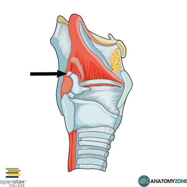
The structure indicated is the corniculate cartilage of the larynx. The corniculate cartilages are also known as the cartilages of Santorini.

The larynx consists of several cartilages, as well as lots of small muscles and a fibroelastic membrane.

There are three pairs of small cartilages, and three large unpaired cartilages.

The large unpaired cartilages include the cricoid cartilage, the thyroid cartilage and the epiglottis.

The small paired cartilages include the arytenoid, the corniculate and the cuneiform cartilages.

The corniculate cartilages are small cone shaped cartilages which sit on the apices of the arytenoid cartilages.

Learn more about the larynx in this tutorial!


Soft Palate

$
0
0

Soft Palate

Soft Palate

The structure indicated is the soft palate.

The hard and soft palates form the roof of the oral cavity. The oral cavity opens anteriorly on the face via the oral fissure. Posteriorly the oral cavity opens into the oropharynx via oropharyngeal isthmus. The floor is formed mainly of the tongue and surrounding soft tissue. Laterally, the oral cavity is bounded by the muscular cheeks.

The hard palate forms the anterior portion of the roof of the oral cavity and consists of a plate of bone (from the maxilla and palatine bones) covered in mucosa.

Posteriorly, the soft palate is continuous with the hard palate. It is called “soft” as the mucosa of which it is formed does not lie over bone, but instead is draped over muscles. There are five muscles which are involved in moving the soft palate:

  1. Tensor veli palatini
  2. Levator veli palatini
  3. Palatopharyngeus
  4. Palatoglossus
  5. Musculus uvulae

The soft palate is mobile and can act to seal off the nasopharynx when it is elevated, and it can be lowered to seal off the oropharyngeal isthmus.

Myenteric Plexus

$
0
0

Myenteric Plexus

Myenteric Plexus

The myenteric plexus, also known as Auerbach’s plexus, together with Meissner’s plexus (submucosal plexus) forms the enteric nervous system which regulates the function of the gastrointestinal system.

The myenteric plexus is located between the inner and outer muscle layers of the muscularis externa. The submucosal plexus (Meissner’s plexus), on the other hand, is located in the submucosa.

This diagram illustrates the four layers of the gastrointestinal tract:

  • Mucosa – consists of 3 layers
    1. Epithelium
    2. Lamina Propria
    3. Muscularis Mucosae
  • Submucosa – connective tissue, submucosal plexus and blood vessels/lymphatics
  • Muscularis Externa – 3 muscle layers in the stomach, 2 in the small/large intestines surrounding the myenteric plexus:
    1. Circular (inner)
    2. Longitudinal (outer)
  • Adventitia/serosa – serous membrane which covers the muscularis externa

The myenteric plexus contains both parasympathetic and sympathetic fibres and supplies the circular and longitudinal muscle layers with motor innervation.

Hirschsprung’s disease is a congenital disorder of the newborn in which ganglion cells of the myenteric plexus are absent due to failure of the cells from the neural crest to migrate into the large intestine as a part of the normal formation of the enteric nervous system during fetal development. The disorder often presents with the baby failing to pass meconium (first stool) following delivery.

Learn more about the anatomy of the autonomic nervous system in this tutorial.

Submandibular Gland

$
0
0

Submandibular Gland

Submandibular Gland

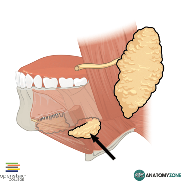
The structure indicated is the submandibular gland.

There are three main large sets of salivary glands in the face:

  1. Parotid glands
  2. Submandibular glands
  3. Sublingual glands

The submandibular glands are hook shaped and consist of a deep part and a superficial part, separated by the mylohyoid muscle.

The deep part of the submandibular gland is formed from the shorter arm of the hook, which loops posteriorly around the mylohyoid muscle to enter the floor of the oral cavity.

The superficial part of the submandibular gland is formed from the longer arm of the hook and lies outside the oral cavity.

The duct of the submandibular gland (Wharton duct) opens adjacent to the inferior point of attachment of the frenulum.

The submandibular glands receive parasympathetic innervation via the chorda tympani branch of the facial nerve (cranial nerve VII).

Seminal Vesicles

$
0
0

Seminal Vesicles

Seminal Vesicles

The structure indicated is the seminal vesicle.

The seminal vesicles are paired accessory glands of the male reproductive system. The seminal vesicles lie lateral to the ductus deferens just beneath the bladder. The excretory duct of the seminal vesicles join the ipsilateral vas deferens to form the ejaculatory ducts. The ejaculatory ducts formed on either side then pass through the prostate to open into the prostatic urethra at the colliculus seminalis.

The seminal vesicles contribute a significant volume of the fluid which forms semen.

Learn about the basic anatomy and structure of the male reproductive system in this anatomy tutorial.

Great Saphenous Vein

$
0
0

Great Saphenous Vein

Great Saphenous Vein

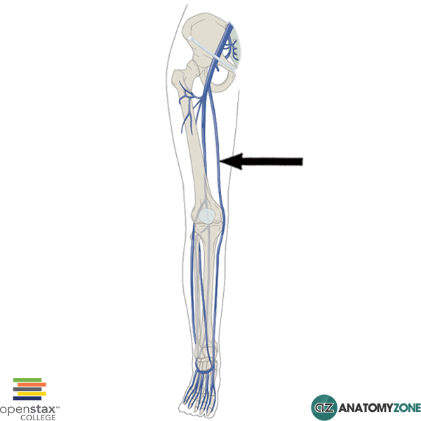
The structure indicated is the great saphenous vein.

The great saphenous vein is a large superficial vein (the longest vein in the human body), which begins from the medial aspect of the dorsal venous arch of the foot. It then passes anteriorly, in front of the medial malleolus and ascends medially up the lower limb to drain into the femoral vein in the femoral triangle.

Learn about the femoral triangle in this anatomy tutorial.

Learn about the veins of the body in this tutorial.

Genioglossus

$
0
0

Genioglossus

Genioglossus

The structure indicated is the genioglossus muscle of the tongue.

The genioglossus muscle is one of the extrinsic muscles of the tongue. The muscles involved with the tongue consist of intrinsic muscles which lie within the tongue itself, and the extrinsic muscles which attach to the tongue and are responsible for depression, elevation, protraction and retraction of the tongue.

There are four sets of extrinsic tongue muscles:

  1. Genioglossus
  2. Hyoglossus
  3. Palatoglossus
  4. Styloglossus

The genioglossus is a large muscle which extends anteriorly from the chin, to the tongue. The genioglossus is involved in depression and protrusion of the tongue.

Origin: Superior mental tubercles

Insertion: Body of hyoid bone and dorsum of tongue

Action: Protrusion and depression of tongue

Innervation:  Hypoglossal nerve (Cranial nerve XII)

Learn all about the anatomy of the cranial nerves and the extrinsic muscles of the tongue and hyoid bone.

Calcaneus

$
0
0

Calcaneus

Calcaneus

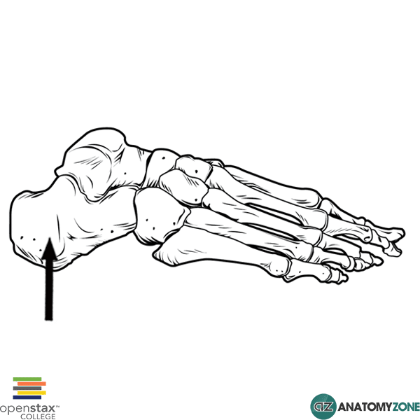
The structure indicated is the calcaneus bone of the foot.

The foot contains three groups of bones:

  • Tarsals
  • Metatarsals
  • Phalanges

The calcaneus is one of seven tarsal bones. There are three groups of tarsal bones:

  • Proximal group
  • Intermediate group
  • Distal group

There are two bones in the proximal group of tarsal bones: the talus and the calcaneus. The calcaneus is the largest tarsal bone and forms the heel through its posterior projection. Anteriorly, the calcaneus articulates with the cuboid bone, and superiorly it articulates with talus.

Three muscles insert onto the calcaneus:

  • Gastrocnemius
  • Soleus
  • Plantaris

Learn more about the bones of the foot in this anatomy tutorial.


Superficial Temporal Artery

$
0
0

Superficial Temporal Artery

Superficial Temporal Artery

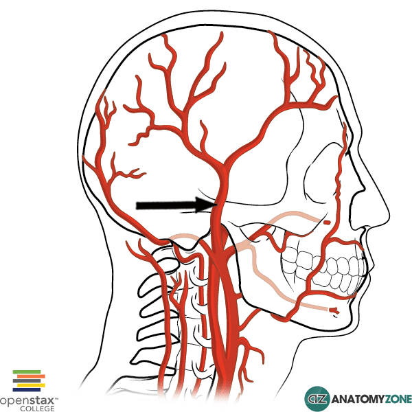
The structure indicated is the superficial temporal artery.

The superficial temporal artery is a branch of the external carotid artery. The external carotid artery gives rise to 8 branches:

  • Superior thyroid artery
  • Ascending pharyngeal artery
  • Lingual artery
  • Facial artery
  • Occipital artery
  • Posterior auricular artery
  • Maxillary artery
  • Superficial temporal artery

The maxillary artery and the superficial temporal artery are terminal branches of the external carotid artery, with the latter being the smaller of the two branches.

The superficial temporal artery is often biopsied when giant-cell arteritis is suspected. The artery can be easily palpated just anterior to the tragus – feel it yourself!

Learn more about the branches of the external carotid artery in this anatomy tutorial.

Omohyoid

$
0
0

Omohyoid

Omohyoid

The structure indicated is the omohyoid muscle.

The omohyoid muscle is one of four infrahyoid muscles which lie in the anterior triangle of the neck. The infrahyoid muscles, as the name suggests, attach to the hyoid bone and lie below it. Contraction of the infrahyoid muscles then causes depression of the hyoid bone. The infrahyoid muscles are often referred to as the strap muscles due to their strap-like appearance.

The four infrahyoid muscles are:

  • Omohyoid
  • Sternohyoid
  • Thyrohyoid
  • Sternothyroid

The omohyoid muscle sits lateral to the sternohyoid muscle and is comprised of two muscle bellies (superior and inferior) connected by a tendon in the middle.

Origin: superior border of scapula

Insertion: lateral to sternohyoid muscle on body of hyoid bone

Action: depression of hyoid bone

Innervation: anterior rami of C1 to C3 (ansa cervicalis)

Learn more about the muscles of the anterior triangle of the neck in this anatomy tutorial.

Abductor Pollicis Brevis

$
0
0

Abductor Pollicis Brevis

Abductor Pollicis Brevis

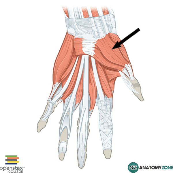
The structure indicated is the abductor pollicis brevis muscle of the hand.

The abductor pollicis brevis muscle is one of the intrinsic muscles of the hand that belongs to the thenar group.

The thenar group of intrinsic hand muscles consist of three muscles:

  1. Abductor pollicis brevis
  2. Flexor pollicis brevis
  3. Opponens pollicis

All the intrinsic muscles of the hand, except the thenar muscles and the lateral two lumbrical muscles are innervated by the deep branch of the ulnar nerve. The thenar muscles and the lateral two lumbrical muscles are innervated by the median nerve.  A useful mnemonic for remembering this is MEATLOAF. “MEAT” refers to the Median nerve, and LOAF refers to the muscles which it innervates: Lateral two lumbricals, Opponens pollicis, Abductor pollicis brevis, Flexor pollicis brevis.

Origin: Flexor retinaculum, tubercles of scaphoid and trapzeium

Insertion: Lateral base of proximal phalanx of thumb

Innervation: Recurrent branch of median nerve: C8, T1

Action: Abducts thumb

Learn all about the anatomy of the muscles of the hand in this tutorial.

Coronary Sinus

$
0
0

Coronary Sinus

Coronary Sinus

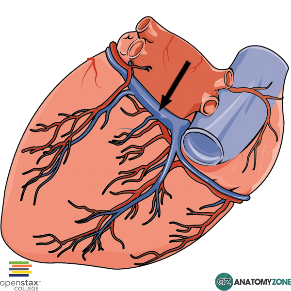
The structure indicated is the coronary sinus.

The coronary sinus lies posteriorly on the heart in the coronary sulcus and receives four cardiac veins:

  1. Great cardiac vein
  2. Middle cardiac vein
  3. Small cardiac vein
  4. Posterior cardiac veins

The coronary sinus returns deoxygenated blood to the right atrium of the heart, together with the superior and inferior vena cavae.

Learn more about the anatomy of the venous drainage of the heart in this tutorial.

Infraspinous Fossa

$
0
0

Infraspinous Fossa

Infraspinous Fossa

The structure indicated is the infraspinous fossa of the scapula.

It is called the infraspinous fossa because it lies below (infra) the spine of the scapula (-spinous). Accordingly, the fossa that lies superior to the spine of the scapula is the supraspinous fossa.

The scapula consists of two surfaces – the costal surface and the posterior surface. The infraspinous fossa is significantly larger than the supraspinous fossa and comprises the majority of the posterior surface of the scapula.

The infraspinatus muscle, one of the rotator cuff muscles, originates in the infraspinous fossa.

Learn all about the anatomy of the rotator cuff muscles and the features of the scapula.

Head of Fibula

$
0
0

Head of Fibula

Head of Fibula

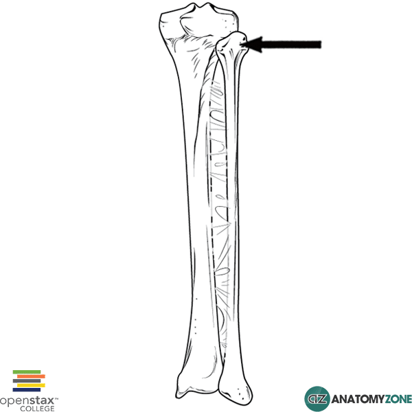
The structure indicated is the head of the fibula.

The two bones of the leg are the tibia and the fibula. The fibula is the smaller of the two bones and lies laterally. The head of the fibula is an expansion of the fibula at the proximal end, and is flattened superiorly and medially forming a surface for articulation with the lateral condyle of the tibia.

The head of the fibula provides the point of attachment for several muscles and ligaments, including the biceps femoris and the lateral collateral ligament (fibular collateral ligament).

Just below the head of the fibula, the common peroneal nerve winds around the neck of the fibula – a point of exposure making it susceptible to damage.

Learn more about the anatomy of the fibula in this tutorial.

Pterion

$
0
0

Pterion

Pterion

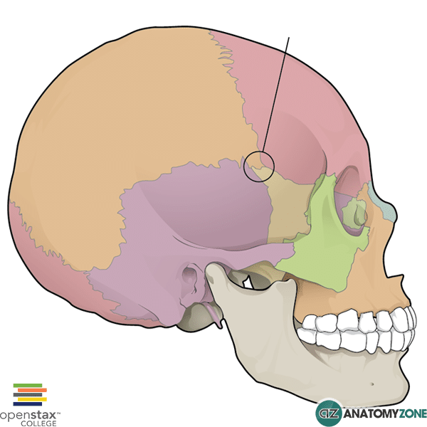
The structure indicated is known as pterion.

Pterion is the name given to the region on the lateral aspect of the skull where four bones are joined:

  • Parietal bone
  • Squamous part of temporal bone
  • Front bone
  • Greater wing of sphenoid bone

Pterion is a point of clinical significance – the skull is very thin at this point. In addition to being structurally weak due to being the point of union between several bones, it also lies over the anterior division of the middle meningeal artery. Fracture of the skull at this point can therefore disrupt the middle meningeal artery, leading to an extradural (epidural) haematoma.

Pterion involves three cranial sutures:

  • Sphenoparietal suture
  • Coronal suture
  • Squamous suture

Learn all about the sutures of the skull in this tutorial.


Infraorbital Foramen

$
0
0

Infraorbital Foramen

Infraorbital Foramen

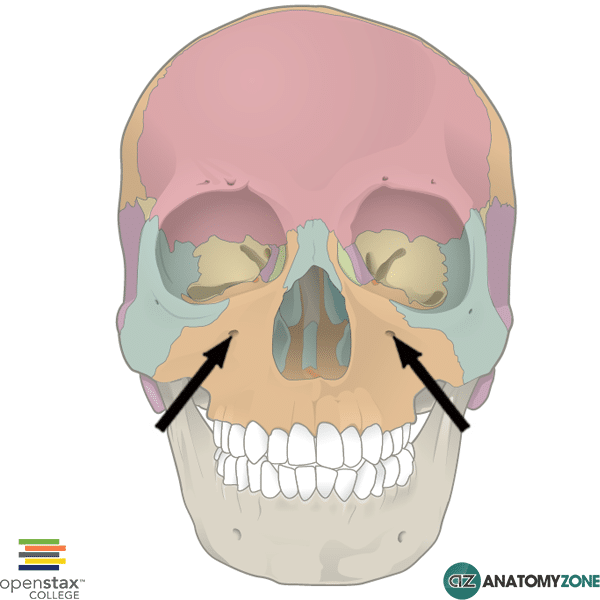
The structure indicated is the infraorbital foramen of the skull.

The infraorbital foramen are a pair of external foramina of the skull which are visible anteriorly on the skull. Foramen (foramina is plural), are holes in the human body which allow other structures to pass through. In the case of the skull, foramina permit the passage of arteries, veins and nerves.

There are three pairs of foramina visible from an anterior view of the skull:

  1. Infraorbital foramen
  2. Supraorbital foramen
  3. Mental foramen

The infraorbital foramen is located in the maxillary bone of the skull just below the inferior margin of the orbit.

The infraorbital foramen permits the passage of three structures:

  1. Infraorbital artery
  2. Infraorbital vein
  3. Infraorbital nerve (a branch of the maxillary division (V2) of the trigeminal nerve [cranial nerve V])

Learn more about the skull and the cranial nerves.

Z-Line

$
0
0

Z-Line

Z-Line

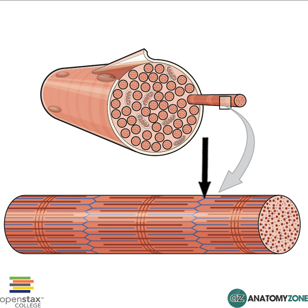
The structure indicated is the Z-disc/Z-line formed between adjacent sarcomeres.

A sarcomere is the name given to the basic unit of muscle, composed of sliding protein filaments of actin and myosin. Myosin filaments are thick, are thin. These two filaments overlap each other, and their ability to slide past each other forms the basis of the theory of muscle contraction and relaxation.

Under electron microscopy, the overlapping pattern of these thick and thin protein filaments gives rise to darker and lighter areas (bands), as well as distinct lines. The thinner actin filaments are all bound to the Z-line, which makes up the boundary of the sarcomere. Z-lines therefore, are found between adjacent sarcomeres, and a sarcomere is thus defined as the muscle unit that is found between Z-lines.

The sarcomere consist of the following bands and zones:

  • I-band – light area around the Z-line consisting of thin actin filaments not overlapped by thick filaments
  • A-band – dark area which spans the length of the thick filament
  • H-zone – light zone in the centre of the a-band where the thick filaments are not overlapped by the thin filaments

The sarcomere consist of two lines:

  • Z-line – forms periphery of sarcomere where thin actin filaments attach
  • M-line – found inside H-zone and forms the middle of the sarcomere.

Anterior Inferior Tibiofibular Ligament

$
0
0

Anterior Inferior Tibiofibular Ligament

Anterior Inferior Tibiofibular Ligament

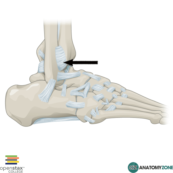
The structure indicated is the anterior inferior tibiofibular ligament.

The distal ends of the tibia and the fibula are held together by the inferior portion of the interosseous membrane. The interosseous membrane is the membrane which joins the tibia to the fibula along its entire length, separating the leg into anterior and posterior compartments. There are two apertures within the interosseous membrane proximally and distally which allow vessels to pass from one compartment to the other.

In addition to the attachment of the interosseous membrane inferiorly between the distal tibia and fibula, this inferior articulation is reinforced by the tibiofibular ligaments anteriorly and posteriorly.

Learn all about the tibia and the fibula in this video tutorial.

Pronator Teres

$
0
0

Pronator Teres

Pronator Teres

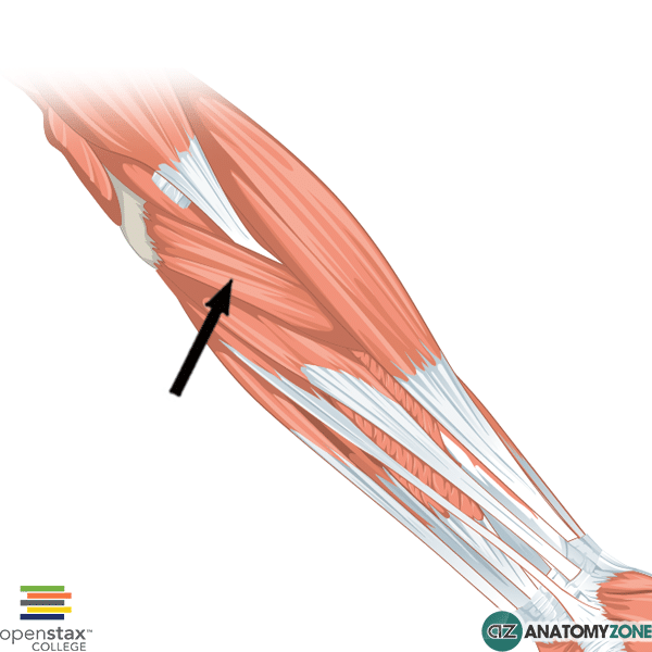
The structure indicated is the pronator teres muscle.

The pronator teres is one of four muscles in the superficial layer of muscles in the anterior compartment of the forearm. The anterior compartment of the forearm consists of three layers of muscles:

  • Superficial layer
  • Intermediate layer
  • Deep layer

Four muscles are located in the superficial layer (from lateral to medial):

  1. Flexor carpi ulnaris
  2. Palmaris longus
  3. Flexor carpi radialis
  4. Pronator teres

All four muscles of the superficial layer have a common origin on the medial epicondyle of the humerus.

Origin:  humeral head – medial epicondyle of humerus; ulnar head – coronoid process

Insertion: middle of lateral radial surface

Action: pronation, weak flexor of elbow

Innervation: median nerve

Learn more about the anatomy of the muscles of the forearm in this tutorial.

Teres Major

$
0
0

Teres Major

Teres Major

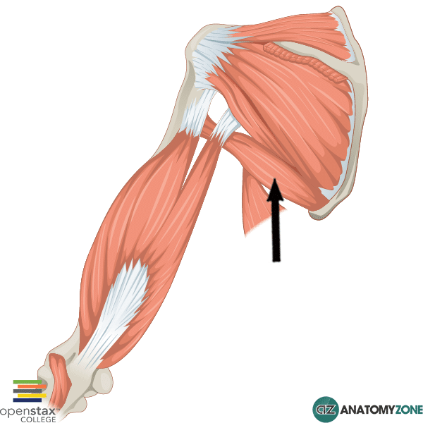
The structure indicated is the teres major muscle.

The teres major muscle is located in the posterior scapular area. It originates on the inferior angle of the scapula and inserts anteriorly on the humerus on the intertubercular sulcus. Contraction of the teres major causes extension and medial rotation of the humerus at the glenohumeral joint.

Origin:  Inferior angle of scapula

Insertion: Medial portion of intertubercular sulcus of humerus

Action: Adduction, medial rotation, extension of humerus

Innervation: Lower subscapular nerve

The pectoralis major inserts on the lateral part of the intertubercular sulcus and the latissimus dorsi inserts on the floor of the intertubercular sulcus. A mnemonic for remembering this pattern of insertion is “The Lady Between Two Majors” – “Lady” referring to “Latissimus dorsi”, and the two majors being the pectoralis major and teres major.

Learn about the features of the scapula in this tutorial

Viewing all 279 articles
Browse latest View live